Introduction
The path of eruption of any tooth can become disturbed. Sometimes the reason is obvious, such as a supernumerary tooth impeding an upper incisor, but often it is obscure. In clinical orthodontics, the most common problem of aberrant eruption is the impacted maxillary canine, which is second only to the third molar in the frequency of impaction.
Prevalence of impacted maxillary canines
Ectopic maxillary canines occur in about 2% of the population, of which about 85% of canines are palatal and 15% buccal to the line of the upper arch. The risk of impaction of the upper canine is greater where the lateral incisor is diminutive or absent¾the lateral incisor root is known to guide the erupting canine. An impacted canine can sometimes resorb adjacent incisor roots, and this risk may be as high as 12%. Incisor resorption is sometimes quite dramatic.

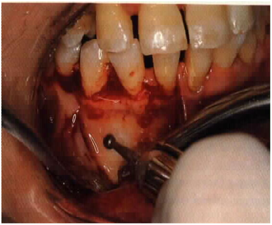
Impacted canine causing root resorption
Clinical
assessment
During the mixed dentition stage the normal path of eruption of the maxillary canines is slightly buccal to the line of the arch, and from about 10 years of age the crowns should be palpable as bulges on the buccal aspect of the alveolus.
If not, an abnormal path of eruption should be suspected, particularly where eruption of one canine is very delayed compared with the other side. Unerupted maxillary canines should be palpated routinely on all children from the age of 10 years until eruption.
Radiographic assessment
Where the canine is not palpable it should be assessed radiographically. A periapical radiograph shows whether the primary canine root is resorbing normally and whether the canine follicle is enlarged. If the apex of the primary canine is not resorbing, with either no root resorption or only lateral resorption, the path of eruption of the permanent canine may be abnormal. However, a single radiograph cannot fully determine the unerupted canine's position relative to the other teeth¾two views are needed for this, either at right angles to each other or for the parallax technique.
Parallax technique
This method, also known as the tube-shift method, compares two views of the area taken with the X-ray tube in two different positions. (a) shows a palatal canine on a periapical film being taken with the tube positioned forward or mesially. A second film taken with the tube positioned further distally gives an image which apparently shows the canine crown in a different position relative to the adjacent roots. In this case the image of the canine appears to have shifted distally when compared with the first film, that is in the same direction that the tube was moved, which indicates that the canine is palatal to the other teeth. An apparent shift in the opposite direction to the tube shift would indicate that the tooth is lying buccally to the other teeth.
The parallax technique works best using two periapical views, but with care it can also be applied to a panoramic tomogram with a standard occlusal view, using vertical shift. The tube position is low down for the panoramic tomogram and much higher for the occlusal view, and so in this example the palatal canine appears to be nearer the incisor apices in the occlusal view, i.e. its apparent movement is upwards with the tube. The size of the image of a displaced tooth on a panoramic radiograph is another indicator, being enlarged if it is palatal and reduced if it is labial or buccal. However, a periapical view is still necessary to check for associated pathology, and this can be used with the occlusal view to make another parallax pair. The combination of panoramic, standard occlusal, and periapical views, such as that in, allows comprehensive assessment of a maxillary canine.
Two films at right angles
This method is more applicable to the specialist as it involves a taking lateral skull view and a posteroanterior (p-a) view: possibly a p-a skull, but more commonly using a panoramic radiograph for the same purpose. The lateral skull view shows whether the canine crown is buccal or palatal to the incisor roots, and the p-a or panoramic view shows how close it is to the mid-line. The angulation of the tooth and its vertical position are assessed using both views. An intraoral view must also be taken to check for any associated pathology.
The position of the impacted canine's crown should be determined as being buccal, palatal, or in the line of the arch. The degree of displacement should be assessed horizontally, that is how close it is to the mid-line, in terms of how far it overlaps the roots of the incisors. The canine crown's vertical position is assessed relative to the incisor apices. An estimate should also be made of the tooth's angulation and the position of its apex relative to the line of the arch.
Other radiographic signs that may suggest an abnormal path of eruption are: obvious asymmetry between the positions of the two upper canines; lack of resorption of the root of the primary canine on the affected side; and resorption of permanent incisor roots. If there are signs of incisor resorption, urgent advice and treatment should be sought.

Parallax location of |3. (a)
Radiograph taken with the tube positioned forward shows that the image of the
canine crown is slightly mesial to the image of |1. (b) Radiograph taken
with the tube positioned further distally shows that the image of |3 is
further distally. The image of |3 has shifted in the same direction as
the tube shift:|3 is therefore nearer to the film than |1, i.e.
it is palatal to the line of the arch. (c) Diagrammatic representation of how a
palatally positioned tooth moves 'with' the tube from left to right

Early
treatment
During the later mixed dentition, if an upper canine is not palpable normally and is found to be ectopic, extraction of the primary canine has a good chance of correcting or improving the path of eruption of the permanent canine, provided it is not too severely displaced. Extraction of the primary canine is only appropriate under these conditions:
(1) early detection¾mixed dentition;
(2) canine crown overlap of no more than half the width of the adjacent incisor root as seen on a panoramic view;
(3) canine crown no higher than the apex of the adjacent incisor root;
(4) angle of 30° or less between the canine's long axis and the mid-sagittal plane;
(5) reasonable space available in the arch¾no more than moderate crowding.
Unless the upper arch is spaced, the contralateral primary canine should also be removed to prevent the upper centreline shifting. Eruption of the permanent canine should be monitored clinically and if necessary radiographically, and specialist advice sought if it fails to show reasonable improvement after a year.
The main disadvantage of extracting the primary canine is losing the option of retaining it if the permanent canine fails to erupt. It may also allow forward drift of the upper buccal teeth where there is a tendency to crowding, and if space is critical a space maintainer should be fitted.
Later treatment
The treatment options in the permanent dentition are to:
(1) expose the canine and align it orthodontically;
(2) transplant the canine;
(3) extract the canine;
(4) leave the impacted canine in situ.
Exposure and orthodontic alignment
This is the treatment of choice for a well-motivated patient, provided the impaction is not too severe. The canine should lie within these limits:
(1) canine crown overlapping no more than half the width of the central incisor root;
(2) canine crown no higher than the apex of the adjacent incisor root;
(3) canine apex in the line of the arch.
The tooth can either be exposed into the mouth and the wound packed open, or a bracket attached to a gold chain can be bonded to it and the wound closed. An orthodontic appliance, usually fixed, then applies traction to bring the tooth into alignment. This treatment can take up to 2 years, depending on the severity of the canine's displacement. Exposure works well for palatally impacted canines, but buccally impacted canines usually have a poor gingival contour following exposure, even when an apically repositioned flap procedure has been used. For this reason some operators prefer to attach a chain to buccally impacted canines and to close the wound, so that the unerupted canine is brought down to erupt through attached, rather than free, gingiva.
Transplantation
The attraction of transplantation is that orthodontic treatment is avoided and yet the canine is brought into function. Two criteria must be met: the canine can be removed intact with a minimum of root handling; and there must be adequate space for the canine in the arch.
The major cause of failure is root resorption, but the incidence of this is reduced if the surgical technique is atraumatic and the transplanted tooth is root- filled with calcium hydroxide shortly after surgery. The success rate for canine transplantation is about 70% survival at 5 years, but many clinicians regard it as being appropriate in only a few cases.
Extraction of the permanent canine
This is appropriate if the position of the canine puts it beyond orthodontic correction, or if the patient does not want appliance treatment. If present, the primary canine can be left in situ, and although the prognosis is unpredictable, a canine with a good root may last for many years. When it is eventually lost a prosthesis will be needed, and provision of this can be difficult if the overbite is deep¾another factor to be taken into account when considering treatment options.
Extraction of the permanent canine may also be considered where the lateral incisor and premolar are in contact, giving a good appearance. In this case it is often expedient to accept the erupted teeth and extract the canine.
Leaving the unerupted canine in situ
During the early teenage years there is a risk of resorption of adjacent incisor roots so that annual radiographic review is necessary, although the risk of root resorption reduces with increasing age. The onset of root resorption can be quite rapid, and for this reason many impacted canines are removed. There may be a case for retaining the canine in the short term in a younger patient, in case they have a change of heart about orthodontic treatment to align the tooth.
Key Points
Ectopic canines
· About 2% of children have ectopic upper canines, of which 85% are palatal.
· Always palpate for upper canines from the age of 10 years until eruption.
· Non-palpable upper canines should be located radiographically or referred for investigation.
· Consider extraction of a primary canine if a permanent canine is mildly displaced.
· Untreated, unerupted permanent canines may resorb incisor roots and should be radiographed annually during the teenage years.