Oral
manifestations of nutritional deficiencies include nonspecific signs
and symptoms that involve the mucous membrane, the teeth and the
periodontal tissues, salivary glands and peri-oral skin. Owing to the
rapid rate of cell turn over in the mucous membrane(3-7 days), compared
with the skin (up to 28 days) the oral cavity may exhibit early signs
and symptoms of systemic disease or nutritional deficiencies. Thus
dentist may be the first person to detect nutritional deficiencies of an
individual and should be able to differentiate it from local causes.
Vitamins are organic components in food that are needed in very small amounts for growth and for maintaining good health.
There are mainly two types of vitamins.
· Fat soluble- which can be stored with other lipids in fatty tissues or in the liver and can build up to toxic levels. Require bile for absorption
Ex. vitamin A (Beta Carotene), vitamin D, vitamin E, vitamin K (Menadione).
· Water soluble - absorbed directly in the blood stream, travel freely and are not stored. Excreted in the urine.
Ex. folate (folic acid), biotin, Vitamin B-1 (Thiamin), Vitamin B-2 (riboflavin), Vitamin B-3 (Niacinamide), vitamin B-5, vitamin B6 (Pyridoxine) vitamin B12 (Cobalamin), pantothenic acid, and vitamin C (ascorbic acid)
Vitamins
are required in the diet in only tiny amounts, in contrast to the
energy components of the diet such as sugars, starches, fats, and oils.
Not all the vitamins affect the oral mucosa. Water soluble vitamins have
oral mucosal involvement include vitamins B-2, B-3, B-6, B-12, folic
acid and vitamin C whereas vitamins A, D, E fat soluble vitamins affect
oral mucosa.
FAT SOLUBLE VITAMINS
Vitamin A
Vitamin
A (retinol) is part of the family of retinoids which is present in food
and the body as esters combined with long-chain fatty acids.

Retinol is stored in the liver and is transported in plasma bound to an α-globulin, retinol-binding protein (RBP).
Function-
- Vision- Retinaldehyde in its cis form is found in the opsin proteins in the rods (rhodopsin) and cones (iodopsin) of the retina. Light causes retinaldehyde to change to its trans isomer, and this leads to changes in membrane potentials that are transmitted to the brain.
- Retinol and retinoic acid are involved in the control of cell proliferation and differentiation.
- Retinyl phosphate is a cofactor in the synthesis of most glycoproteins containing mannose.
reference nutrient intake (RNI)- 700 μg
lower reference nutrient intake (LRNI)- 300 μg
Deficiency-
 |
| keratosis of labial mucosa |
Inadequate
cell differentiation-impaired healing & tissue regeneration;
desquamation of oral mucosa; keratosis; increased risk of candidiasis;
gingival hypertrophy & inflammation; leukoplakia; decreased taste
sensitivity; xerostomia; disturbed or arrested enamel development;
irregular tubular dentine formation and increased caries risk.
Other - Xerophthalmia, night blindness, keratomalacia, follicular hyperkeratosis.
 |
| xerophthalmia |
Diagnosis –
· Clinical diagnosis where nutritional deficiencies common
· Blood vit.A level
· Response to replacement therapy
Treatment-
· retinol palmitate 30 mg orally for two days
· treat associated malnutrition and super added infection
Adverse effects-
- High intakes of vitamin A-Chronic ingestion of retinol can cause liver and bone damage, hair loss, double vision, vomiting, headaches and other abnormalities. Single doses of 300 mg in adults or 100 mg in children can be harmful.
- Retinol is teratogenic-The incidence of birth defects in infants is high with vitamin A
Vitamin D
active form 1,25-dihydroxycholecalciferol (1,25-(OH)2D3).
The
primary source of vitamin D in humans is photoactivation (in the skin)
of 7-dehydrocholesterol to cholecalciferol, which is then converted in
the liver to 25-hydroxycholecalciferol (25-(OH)D3) and further converted by renal 1α-hydroxylase to the active metabolite.

lower reference nutrient intake (LRNI)- 10 μg (living indoors)
Function-
· Maintain serum calcium level within normal range
Deficiency-
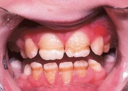 |
| enamel hypoplasia |
General-
Causes hypocalcaemia, which leads to excess parathyroid hormone
secretion, causing bone demineralization. Which leads to,
 |
| vitamin D dependent Rickets |
· Osteomalacia
Diagnosis-
· Serum calcium and phosphate level-low
· Plasma alkaline phosptasw level-high
· 1,25-dihydroxycholecalciferol levels( ≥20 ng/mL considered normal)
· X ray wrist joint in rickets
Treatment-
· Advice on balanced diet
· Correction of predisposing factors
· Daily administration of Vit.D3
Vitamin E
Divided in to mainly two compounds.
· Tocopherols-commonest is α-tocopherol
· Tocotrienoles
Sources- Vegetables and seed oils, including soya bean, saffron, sunflower, cereals and nuts

· Act as a antioxidant
· affect cell proliferation and growth
deficiency-
no oral manifestations apperant.
· severe neurological deficits (gross ataxia)
· severe neurological deficits (gross ataxia)
· aging effects on skin, hair, nails etc.
diagnosis-
· plasma α-tocopherol level corrected for the level of plasma lipids ( value as per milligram of plasma lipid or cholesterol)
treatment-
· vit E injection
· vit E capsules
Vitamin K
Vitamin K is found as phylloquinone (vitamin K1) and menaquinone (vitamin K2)
Source- K1- green leafy vegetables, dairy products, rape seed and soya bean oils
K2-Intestinal bacteria synthesizes in the terminal ileum and colon.
reference nutrient intake (RNI)- 1 μg/kg
Function-
· A cofactor for the production of blood clotting factors.
for
the post-translational carboxylation of specific protein-bound
glutamate residues in γ-carboxyglutamate (Gla). Gla residues bind
calcium ions to phospholipid templates, and this action on factors II,
VII, IX and X, and on proteins C and S, is necessary for coagulation
· for proteins necessary in the formation of bone.
Bone
osteoblasts contain three vitamin K-dependent proteins, osteocalcin,
matrix Gla protein and protein S, which have a role in bone matrix
formation. Osteocalcin contains three Gla residues which bind tightly to
the hydroxyapatite matrix depending on the degree of carboxylation;
this leads to bone mineralization.
Deficiency-
oral manifestations-Increased risk of bleeding & candidiasis
other-· increase in the prothrombin time and haemorrhage
causes-
o Cholestatic jaundice
o vitamin K antagonists
o Oral anticoagulants- ex: warfarin
o Certain anti bacterial drugs
WATER-SOLUBLE VITAMINS
Thiamin (vitamin B1)
consists of pyrimidine and thiazole rings. The alcohol side-chain is esterified with one, two or three phosphates.
Source-cereals, grains, beans, nuts, pork and duck
reference nutrient intake (RNI)- 0.4 mg per 1000 kcal of energy requirement
lower reference nutrient intake (LRNI)- 0.23 mg per 1000 kcal
Thiamin diphosphate, often called thiamin pyrophosphate (TPP), is a cofactor in carbohydrate metabolism.
Thiamin deficiency-
no oral manifestations.
· beriberi(Dry beriberi, Wet beriberi)
· Wernicke-Korsakoff syndrome(dementia, ataxia, varying ophthalmoplegia and nystagmus)
causes
- in beriberi, where the only food consumed is polished rice
- in chronic alcohol-dependent patients who are consuming virtually no food at all
- rarely in starved patients
Diagnosis-
· clinical diagnosis in endemic areas
· response to treatment with thiamine
· circulating thiamin concentration
· transketolase activity in red cells using fresh heparinized blood.
Treatment-
· In beri beri- Thiamine 50 mg i.m. is given for 3 days, followed by 25 mg of thiamine daily by mouth.
· In
Wernicke-Korsakoff syndrome- Urgent treatment with thiamine 250 mg i.m.
or i.v. twice daily is given for 3 days combined with other B-complex
vitamins
Riboflavin(Vitamin B2)
Riboflavin is a flavoprotein.
Sources- dairy products, fat and leafy vegetables
reference nutrient intake (RNI)- 1.3 mg

Function-
· cofactor for many oxidative reactions in the cell.
Deficiency-

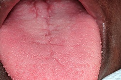 |
| magenta coloured tongue |
 |
| atrophy of filliform papillae |
general-seborrhoeic dermatitis, particularly involving the face (around the nose) and the scrotum or vulva.
Treatment-
Diagnosis-
· In endemic areas mainly a clinical diagnosis
· Riboflavin 5 mg daily usually given as the vitamin B complex

Exist in two chemical forms, nicotinic acid and nicotinamide.
Sources- plants, meat (particularly offal) and fish.
Synthesize in body using tryptophan. Eggs and cheese contain tryptophan
reference nutrient intake (RNI)- 6.6 mg per 1000 kcal
lower reference nutrient intake (LRNI)- 4.4 mg per 1000 kcal
Function-
· act
as hydrogen acceptors in many oxidative reactions, and in their reduced
forms (NADH and NADPH) act as hydrogen donors in reductive reactions.
Deficiency-
oral manifestations-Angular
cheilosis; mucositis; stomatitis; oral pain; ulceration; ulcerative
gingivitis; denuded tongue; glossitis; glossodynia; tip of tongue is red
& swollen; dorsum is dry & smooth.
general-Pellagra,dermatitis, diarrhoea and dementia
Diagnosis-
 |
| pellagra |
 |
| atrophic glossitis in pellagra |
· In endemic areas mainly a clinical diagnosis
Treatment
· Nicotinamide
(approximately 300 mg daily by mouth) with a maintenance dose of 50 mg
daily is given with dramatic improvement in the skin and
diarrhoea.mostly vitamin B complex is given, as other deficiencies are
often present.
· increase in the protein content of the diet.
Vitamin B6
exists as pyridoxine, pyridoxal and pyridoxamine.
Sources- plant and animal foodstuffs.
reference nutrient intake (RNI)- 15 μg per g of dietary protein

Function-
· Pyridoxal phosphate is a cofactor in the metabolism of many amino acids.
Deficiency-
oral manifestations-Angular cheilosis,sore or burning mouth,glossitis,glossodynia.
general-Polyneuropathy
Some drugs (e.g. isoniazid, hydralazine and penicillamine) interact with pyridoxal phosphate, producing B6 deficiency.
Treatment-
Vitamin B complex or Vitamin B6
Vitamin B12
Cobalamins consist of a planar group with a central cobalt atom (corrin ring) and a
right-angles . Vitamin B12
was first crystallized as cyanocobalamin, but the main natural
cobalamins have deoxyadenosyl-, methyl- and hydroxocobalamin groups
attached to the cobalt atom.

reference nutrient intake (RNI)- 1.5 μg
lower reference nutrient intake (LRNI)- 1.0 μg
Function-
· Co
factor in DNA synthesis-methylation of homocysteine to methionine with
the demethylation of methyl THF polyglutamate to THF. THF is a substrate
for folate polyglutamate synthesis.
Absorption and transport-
Vitamin B12 is liberated from protein complexes in food by gastric enzymes and then binds to a vitamin B12-binding protein ('R' binder) related to plasma transcobalamin I (TCI), derived from saliva. Vitamin B12 bound to 'R' binder is released by pancreatic enzymes and becomes bound to intrinsic factor.
Intrinstic factor carries it to specific receptors on the surface of the mucosa of the ileum. Vitamin B12 enters the ileal cells and intrinsic factor remains in the lumen. Vitamin B12 is transported from the enterocytes to the bone marrow and other tissues by the glycoprotein transcobalamin II (TCII).
Deficiency-
 |
| aphthous ulceration |
general- Megaloblastic anemia and Pancytopenia
Sub acute combined degeneration of spinal cord
Causes-
o Low dietary intake
o Vegans
o Impaired absorption-stomach(Pernicious anaemia, Gastrectomy,)Small bowel(Ileal disease or resection, Bacterial overgrowth)
o Congenital transcobalamin II deficiency
o Nitrous oxide (inactivates B12)
o Abnormal utilization
Diagnosis-
· FBC,Blood picture(Macrocytic anemia)
Bone marrow shows the typical features of megaloblastic erythropoiesis
· Serum vitamin B12 is below 160 ng/L
· Serum folate level is normal or high, and the red cell folate is normal or reduced
· Vitamin B12 absorption tests- Schilling test
Treatment
· Hydroxocobalamin
1000 μg can be given intramuscularly to a total of 5-6 mg over the
course of 3 weeks; 1000 μg is then necessary every 3 months.
Folate
Folic acid present in nature as polyglutamates.

lower reference nutrient intake (LRNI)- 100 μg
Function-
· act as coenzymes in the transfer of single carbon units in amino acid metabolism and DNA synthesis.
Deficiency-
oral manifestations-Angular
cheilosis; mucositis; stomatitis; sore or burning mouth; increased risk
of candidiasis; inflamed gingiva; glossitis oral pain; ulceration;
ulcerative gingivitis; denuded tongue; glossitis; glossodynia; tip or
borders of tongue red & swollen; slick bald pale; apthous ulcers.
general- Megaloblastic anemia and Pancytopenia
Causes-
o Nutritional-poor intake(Starvation,alcohol intake,GI diseases )Antifolate drugs( Anticonvulsants, Methotrexate, Pyrimethamine) Malabsorption
o Excess utilization- Physiological(Pregnancy, Lactation)Pathological(haemolysis, Malignant, Inflammatory diseases)
Diagnosis-
· FBC and Blood picture-Macrocytic Anemia,Pancytopenia
· Serum and red cell folate level
Treatment-
· 5 mg of folic acid daily
Vitamin C
Ascorbic acid is a simple sugar.

easily leached out of vegetables when they are placed in water and it is also oxidized to dehydro-ascorbic acid during cooking or exposure to copper or alkalis.
reference nutrient intake (RNI)- 40 mg
lower reference nutrient intake (LRNI)- 10 mg
Function-
· powerful reducing agent, main role is to control the redox potential within cells.
· hydroxylation of proline to hydroxyproline, which is necessary for the formation of collagen
· in high dosage (1-2 g daily) prevention of the common cold
· preventive effect in atherosclerosis and cancer
Deficiency-
 |
| inflamed bleeding gingivae |
general-scurvy- Keratosis of hair follicles with 'corkscrew' hair, Perifollicular haemorrhages, Swollen, spongy gums with bleeding and superadded infection, loosening of teeth,
· and hemorrhages, Anaemia, Failure of wound healing
Diagnosis-
· Hypochromic anemia
· Low Plasma ascorbic acid
· Low leucocyte ascorbate
Treatment-
· encourage to eat fresh fruit and vegetables.


