Important definitions
Malformation definition
A morphologic defect of an organ, part of an organ or large
area of the body resulting from a developmental abnormality (intrinsic).
Eg: Cleft lip
Deformation definition
Abnormal form of the body part due to mechanical forces.
Disruption definition
Defect of an organ, part of an organ or large area of the
body due to interference with a normal developmental process.
Eg: Amneotic bands leads to amputation
Sequence
Multiple defects occur as a result of single presumed
structural abnormality.
Eg: Pierre Robin’s Sequence
Syndrome
Combination of signs and/or symptoms that forms as a
distinct clinical picture indicative of a particular disorder.
Eg: Down’s syndrome
Common Syndromes and
developmental anomalies found in Head and Neck region
- Cleft lip and palate
- Valocardiofacial syndrome
- Pierre Robin’s syndrome
- Treacher Collins syndrome
- Golden har syndrome
- Apert and Crouzen syndrome
Approch to diagnosis
There are over 3000 known syndromes. But only very few are
found in common during dental practice. idea of this post is to explain important
clinical features of commonly found syndromes and their implication on dental
practice.
History plays a major role in diagnosis of a developmental anomaly.
Under history these factors are of uttermost important in diagnosis.
- Medical pedigree
- Maternal and paternal age
- Consanguinity
- Previous abortions
- Teratogens (Fetal alcohol syndrome)
These factors should be thoroughly examined during physical
examination
- Compare siblings and other family member’s photographs
- Major and minor abnormalities
- Isolated minor anomalies (15%)
- More than 3 minor anomalies with major anomalies (90%)
- Mental retardation associated
Down’s syndrome
Etiology: nondisjunction mutation resulting in trisomy of 21
chromosome.
Prevalence 1:700
Down’s syndrome is the most common chromosomal abnormality.
It is associated with maternal age more than 37 years.
Facial characteristics
- Macroglossia
- Micrognathia
- Midface hypoplasia (Class III)
- Flat occiput
- Flat nasal bridge
- Epicanthal fold
- Up slanting palpebral fissures
- Progressive enlargement of lips
Airway concerns
Due to midface hypoplasia
Obstructive sleep apnoea
Prevalence: 54-100% down’s patients.
As a combination of
anatomic and functional mechanisms.(midface hypoplasia,macroglossia, etc. and
hypotonia of pharyngeal muscles)
Cardiovascular abnormalities found in 40% down’s patints.
ASD,VSD, Tetralogy of fallot’s, PDA
Gastrointestinal abnormalities found in 10-18% of down’s
patients.
Malignancies are associated with some down’s cases
Valocardiofacial Syndrome (VCFS)
Autosomal dominant condition. Velocardiofacial syndrome
(VCFS) is a genetic condition characterized by abnormal pharyngeal arch development
that results in defective development of the parathyroid glands, thymus, and
conotruncal region of the heart.
Etiology: by deletion
of chromosome 22
Clinical Features: congenital heart diseases, hyper nasal
speech, and cleft palate
Basicranial angle flexion- Therefore longer face
Puffy eyelids
Vascular anomalies are very common
Anomalies of the head and neck region are common.
Treacher collin’s syndrome
Autosomal dominant condition
60% are from new mutation
Characteristics: likely due to abnormal migration of neural
crest cells into first and second branchial arch structures.
Usually bilateral and symmetrical
Down slanting eyebrows and palpebral fissures
Retruded mandible
Apert and Crowzen’s syndromes
Belong to family of craniosynostosis
Apert’s syndrome is also called as Acrocephalosyndactyly
Facial Characteristics:
- Craniosynostosis (coronal sutures fused at birth and head circumference is larger than the average head circumference.
- Hypertelorism
- Shallow orbits with Exopthalmus
- Maxillary hypoplasia causing retruded midface with relative prognathism.
- Beaked nose
- Downward slanting palpebral fissures
- Syndactyly
Airway concerns
Treatment
Fronto orbital advancement surgery can be done and get good
protection to eyes. Syndactyly can be corrected with surgical reconstruction.
Pierre Robin’s syndrome
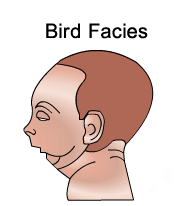
Characterize by triad of micrognathia, Glossoptosis, Cleft
palate.
80% syndromic
Mechanism of cleft palate formation
Mandibular deficiency causes hypoplastic and retruded
mandible (Micrognathia). Therefore Tongue remains retruded and high in
oropharynx causing failure of fusing lateral palatal shelves. This results in
cleft palate.
Airway obstruction is a marked feature.
Pierre Robin’s syndrome can be associated with other
anatomic and neuromuscular components.
Airway management
Temporizing modalties
Prove positioning
Nasopharyngeal airway
Mandibular traction devices
Tongue lip adhesion.
Tracheostomy done if above not worked
Distraction osteogenisis can be done in infants
Goldenhar syndrome
Features
Facial asymmetry and ear deformity.
Vertebral malformations
Uppereyelid coloborma
Auricular malformations
EAC stenosis
Vascular anomalies during fetal life
Unilateral craniofacial malformations
1st and 2nd arch defects
Other concerns
Hearing concerns in greater than 50% patients
Sensoneural occationally.


No comments:
Post a Comment