Friday, July 15, 2011

Odontogenic Infections-Oral Surgery Lecture Note

In order to understand how odontogenic infections are treated, the dentist must be familiar with the terminology concerning infection and the pathophysiology of inflammation, which are described below.
Inoculation
is characterized by the entry of pathogenic microbes into the body without disease occurring.An infection involves the proliferation of microbes resulting in triggering of the defense mechanism, a process manifesting as inflammation.
Inflammation
is the localized reaction of vascularand connective tissue of the body to an irritant, resulting in the development of an exudate rich in proteins and cells. This reaction is protective and aims at limiting or eliminating the irritant with various procedures while the mechanism of tissue repair is triggered. Depending on the duration and severity, inflammation is distinguished as acute, subacute or chronic.
Acute Inflammation
This is characterized by rapid progression and is associated with typical signs and symptoms. If it does not regress completely, it may become subacute or chronic.
Subacute Inflammation
This is considered a transition phase between acute and chronic inflammation.
Chronic Inflammation
This procedure presents a prolonged time frame with slight clinical symptoms and is characterized mainly by the development of connective tissue. Inflammation may be caused by, among other things, microbes, physical and chemical factors, heat, and irradiation. Regardless of the type of irritant and the location of the defect, the manifestation of inflammation is typical and is characterized by the following clinical signs and symptoms: rubor (redness), calor (heat), tumor (swelling or edema), dolor (pain), and functio laesa (loss of function).
The natural progression of inflammation is distinguished into various phases. Initially vascular reactions
with exudate are observed (serous phase), and then the cellular factors are triggered (exudative or cellular phase). The inflammation finally resolves and the destroyed tissues are repaired. On the other hand, chronic inflammation is characterized by factors of reparation and healing. Therefore, while acute inflammation is exudative, chronic inflammation is productive (exudative and reparative). Understanding the differences between these types of inflammation is important for therapeutic treatment. Serous Phase. This is a procedure that lasts approximately 36 h, and is characterized by local inflammatory edema, hyperemia or redness with elevated temperature, and pain. Serous exudate is observed at this stage, which contains proteins and rarely polymorphonuclear leukocytes.
Cellular Phase.
This is the progression of the serous phase. It is characterized by massive accumulation of polymorphonuclear leukocytes, especially neutrophil granulocytes, leading to pus formation. If pus forms in a newly developed cavity, it is called an abscess. If it develops in a cavity that already exists, e.g., the maxillary sinus, it is called an empyema.
Reparative Phase
During inflammation, the reparative phenomena begin almost immediately after inoculation. With the reparative mechanism of inflammation, the products of the acute inflammatory reaction are removed and reparation of the destroyed tissues follows. Repair is achieved with development of granulation tissue, which is converted to fibrous connective tissue, whose development ensures the return of the region to normal.
Infections of the Orofacial Region
The majority (i.e., 90–95%) of infections that manifest in the orofacial region are odontogenic. Of these, approximately 70% present as periapical inflammation, principally the acute dentoalveolar abscess, with the periodontal abscess following, etc.
Periodontal abscess originating from a maxillary central incisor.
Radiograph of same case showing bone resorption, which led to the formation of a periodontal pocket
Etiology
The cardinal causes of orofacial infections are non-vital teeth, pericoronitis (due to a semi-impacted mandibular tooth), tooth extractions, periapical granulomas that cannot be treated, and infected cysts. Rarer causes include postoperative trauma, defects due to fracture, salivary gland or lymph node lesions, and infection as a result of local anesthesia.
Periodontal Abscess
This is an acute or chronic purulent inflammation, which develops in an existing periodontal pocket. Clinically, it is characterized by edema located at the middle of the tooth, pain, and redness of the gingiva. These symptoms are not as severe as those observed in the acute dentoalveolar abscess, which is described below.
Treatment of the periodontal abscess is usually simple and entails incision, through the gingival sulcus with a probe or scalpel, of the periodontal pocket that has become obstructed. Incision may also be performed at the gingivae; more specifically, at the most bulging point of the swelling or where fluctuation is greatest.
Acute Dentoalveolar Abscess
This is an acute purulent inflammation of the periapical tissues, presenting at nonvital teeth, especially when microbes exit the infected root canals into periapical tissues. Clinically, it is characterized by symptoms that are classified as local and systemic.
Local Symptoms
Pain
The severity of the pain depends on the stage of development of the inflammation. In the initial phase the pain is dull and continuous and worsens during percussion of the responsible tooth or when it comes into contact with antagonist teeth. If the pain is very severe and pulsates, it means that the accumulation of pus is still within the bone or underneath the periosteum. Relief of pain begins as soon as the pus perforates the periosteum and exits into the soft tissues.
Edema
Edema appears intraorally or extraorally and it usually has a buccal localization and more rarely palatal or lingual. In the initial phase soft swelling of the soft tissues of the affected side is observed, due to the reflex neuroregulating reaction of the tissues, especially of the periosteum. This swelling presents before suppuration, particularly in areas with loose tissue, such as the sublingual region, lips, or eyelids. Usually the edema is soft with redness of the skin. During the final stages, the swelling fluctuates, especially at the mucosa of the oral cavity. This stage is considered the most suitable for incision and drainage of the abscess.
Other Symptoms
There is a sense of elongation of the responsible tooth and slight mobility; the tooth feels extremely sensitive to touch, while difficulty in swallowing is also observed.
Systemic Symptoms
The systemic symptoms usually observed are: fever, which may rise to 39–40 °C, chills,malaise with pain inmuscles and joints, anorexia, insomnia, nausea, and vomiting. The laboratory tests show leukocytosis or rarely leukopenia, an increased erythrocyte sedimentation rate, and a raised C-reactive protein (CRP) level.
Complications
If the inflammation is not treated promptly, the following complications may occur: trismus, lymphadenitis at the respective lymph nodes, osteomyelitis, bacteremia, and septicemia.
Diagnosis
Diagnosis is usually based upon clinical examination and the patient’s history. What mainly matters, especially in the initial stages, is the localization of the responsible tooth. In the initial phase of inflammation, there is soft swelling of the soft tissues. The tooth is also sensitive during palpation of the apical area and during percussion with an instrument, while the tooth is hypermobile and there is a sense of elongation. In more advanced stages, the pain is exceptionally severe, even after the slightest contact with the tooth surface. Tooth reaction during a test with an electric vitalometer is negative; however, sometimes it appears positive,which is due to conductivity of the fluid inside the root canal.
Radiographically, in the acute phase, no signs are observed at the bone (whichmay beobserved 8–10 days later), unless there is recurrence of a chronic abscess,where upon osteolysis is observed. Radiographic verification of a deeply carious tooth or restoration very close to the pulp, as well as thickening of the periodontal ligament, are data that indicate the causative tooth.
Differential diagnosis of the acute dentoalveolar abscess includes the periodontal abscess, and the dentist must be certain of his or her diagnosis, because treatment between the two differs.
Spread of Pus Inside
Tissues From the site of the initial lesion, inflammation may spread in three ways:
1. By continuity through tissue spaces and planes.
2. By way of the lymphatic system.
3. By way of blood circulation.
Diagrammatic illustrations showing spread of infection (propagation of pus) of an acute dentoalveolar abscess,depending on the position of the apex of the responsible tooth. a Buccal root: buccal direction.b Palatal root: palatal direction
The most common route of spread of inflammation is by continuity through tissue spaces and planes and usually occurs as described below. First of all, pus is formed in the cancellous bone, and spreads in various directions by way of the tissues presenting the least resistance.
a Spread of pus towards the maxillary sinus, due to the closeness of the apices to the floor of the antrum.b Diagrammatic illustration showing the localization of infection above or below the mylohyoid muscle, depending on the position of the apices of the responsible tooth
Whether the pus spreads buccally, palatally or lingually depends mainly on the position of the tooth in the dental arch, the thickness of the bone, and the distance it must travel.Purulent inflammation that is associated with apices near the buccal or labial alveolar bone usually spreads buccally, while that associated with apices near the palatal or lingual alveolar bone spreads palatally or lingually respectively. For example,the palatal roots of the posterior teeth and the maxillary lateral incisor are considered responsible for the palatal spread of pus, while the mandibular third molar and sometimes the mandibular second molar are considered responsible for the lingual spread of infection. Inflammation may even spread into the maxillary sinus when the apices of posterior teeth are found inside or close to the floor of the antrum. The length of the root and the relationship between the apex and the proximal and distal attachments of various muscles also play a significant role in the spread of pus. Depending on these relationships, in the mandible pus originates from the apices found above themylohyoid muscle, and usually spreads intraorally, mainly towards the floor of the mouth (sublingual space). When the apices are found beneath the mylohyoid muscle (second and third molar), the pus spreads towards the submandibular space, resulting in extraoral localization. Infection originating from incisors and canines of the mandible spreads buccally or lingually, due to the thin alveolar bone of the area. It is usually localized buccally if the apices are found above the attachment of the mentalis muscle. Sometimes, though, the pus spreads extraorally, when the apices are found beneath the attachment. In the maxilla, the attachment of the buccinators muscle is significant.When the apices of the maxillary premolars and molars are found beneath the attachment of the buccinator muscle, the pus spreads intraorally; however, if the apices are found above its attachment, infection spreads upwards and extraorally. Exactly the same phenomenon is observed in the mandible as in the maxilla if the apices are found above or below the attachment of the buccinator muscle.
Spread of pus depending on the length of root and attachment of buccinators muscle. a Apex above attachment: accumulation of pus in the buccal space. b Apex beneath the buccinator muscle: intraoral pathway towards the mucobuccal fold
In the cellular stage, depending on the pathway and inoculation site of the pus, the acute dentoalveolar abscessmay have various clinical presentations, such as:(1) intraalveolar, (2) subperiosteal, (3) submucosal, (4) subcutaneous, and (5) fascial ormigratory – cervicofacial.

Intraalveolar abscess of maxilla (a) and mandible (b). Diagrammatic illustrations show accumulation of pus at a portion of the alveolar bone in relation to the periapical region
The initial stage of the cellular phase is characterized by accumulation of pus in the alveolar bone and is termed an intraalveolar abscess . The pus spreads outwards from this site and, after perforating the bone, spreads to the subperiosteal space, from which the subperiosteal abscess originates, where a limited amount of pus accumulates between the bone and periosteum. After perforation of theperiosteum, the pus continues to spread through the soft tissues in various directions. It usually spreads intraorally, spreading underneath the mucosa forming the submucosal abscess.
Subperiosteal abscess with lingual localization. a Diagrammatic illustration; b clinical photograph
Sometimes, though,it spreads through the loose connective tissue and, after its pathway underneath the skin, forms a subcutaneous abscess, while other times it spreads towards the fascial spaces, forming serious abscesses called fascial space abscesses.
Subcutaneous abscess originating from a mandibular tooth. a Diagrammatic illustration. b Clinical photograph.The swelling mainly involves the region of the angle of the mandible
The fascial spaces are bounded by the fascia, which may stretch or be perforated by the purulent exudate, facilitating the spread of infection. These spaces are potential areas and do not exist in healthy individuals, developing only in cases of spread of infection that have not been treated promptly. Some of these spaces contain loose connective tissue, fatty tissue, and salivary glands,while others contain neurovascular structures. Acute diffuse infection,which spreads into the loose connective tissue to agreat extent underneath the skin with or without suppuration,is termed “cellulitis” (phlegmon), and is described in next posts.
Fascial abscess (submandibular). a Diagrammatic illustration. b Clinical photograph
Next Post : Fundamental principles of treatment of infection
Tags : Odontogenic infections, Infections of Oromaxillofacial Region, Infection from lower 3rd molar,Spread of infections,Factors determining spread of infections,Spread of Infections.

Fundamental Principles of Treatment of Infection-Oral Surgery Lecture 

Key Words : oral surgeons wisdom teeth extraction dental implant surgery doctor of dental surgery oral surgeon dental operation dental gum surgery wisdom tooth surgery dental surgery oral surgery doctor of dental surgery cosmetic dental surgery dental laser surgery cosmetic gum surgery laser gum surgery


Monday, July 11, 2011

Elastic Impression Materials-Elastomers-Prosthetic Dentistry Lecture Note

Introduction to Elastic Impression Materials

These materials can be stretched and bent to a fairly large degree without suffering any deformation. These are used for recording the patient's mouth where undercuts are present. Usually used for partial dentures, over dentures, implants and crown and bridge work .The elastic impression materials are:

Introduction to Elastomers

These are used where a high degree of accuracy is needed, especially in crown and bridge work. They have two main advantages over the Hydrocolloids - good tear resistance and dimensional stability.They are mainly hydrophobic rubber based materials. All of these materials come in different viscosity's ranging from low to high viscosity. The light bodied material maybe used as a wash impression over a medium or heavy-bodied material. There are two ways this can be carried out as described below.

ONE STAGE IMPRESSION

Light bodied impression material is placed in a syringe, and placed over the areas where high detail is required (e.g. over a crown preparation). Some is then squirted over the heavy-bodied impression material which has been loaded into an impression tray. The impression is then taken as normal. This technique saves time, but it can be very labour intensive because the two need to mixed at the same time often requiring more than one DSA.

TWO STAGE IMPRESSION

An impression is taken with the heavy-bodied material. This is then removed from the mouth and inspected. The light bodied material is then prepared and again placed in a syringe. This is then squirted over heavy-bodied material and then impression relocated in its original impression.

Polysulphides

Silicones

Polyethers

Polysulphides

CLINICALLY

Used for crown and bridge work mainly, but also used for partial dentures, overdentures and implants. Two equal lengths are mixed together with a spatula for about a minute. The tray needs to be treated with an adhesive (rubber solution in acetone) to provide retention for the polysulphide. Taking the impression is delayed by 5 minutes before the impression is placed in the patients mouth - the final setting time is usually about 10 minutes from the start of mixing - this delay therefore decreases the amount of time the impression tray is in the patients mouth. A one or two stage impression technique may be used. Although dimensionally stable, the impression should be cast within 24 hours.

CHEMISTRY

Other names : Thiokol rubbers, rubber base or mercaptan.
Supplied as two pastes mixed in a 1:1 ratio.

BASE PASTE

· Polysulphide (forms rubber on polymerisation)

· Filler (to give body)

· Plasticiser (control viscosity)

ACTIVATOR PASTE

· Inert oil (forms a paste)

· Sulphur (facilitates the reaction)

· Lead oxide (causes polymerisation and cross-linking)

The active constituent in the base paste is the polysulphide and the active constituents in the activator paste is lead oxide and sulphur which cause further polymerisation of the polysulphide.
On mixing crosslinking and chain lengthening causes the material to become an elastic solid after about 5-8 minutes. Setting is more rapid in the presence of moisture.
They come in three types: light, regular and heavy bodied (viscosity increasing from light through to heavy bodied). The light bodied polysulphides are used as wash impressions on heavier bodied impression materials. The medium and heavy-bodied impression materials may be used on their own.

PROPERTIES

  • Dimensional stability
  • Excellent surface detail (is only used in special trays)
  • Viscosity depends on the brand used
  • Very small setting contraction (0.3-0.4% over the first 24 hrs)
  • Contraction on cooling from mouth to room temperature
  • Very good tear resistance
  • Good shelf life
  • Viscoelastic

ADVANTAGES

  1. Dimensional stability
  2. Accuracy
  3. Comes in a number of different viscosity's
  4. Long working time (although this may be a disadvantage in some clinical situations)
  5. Long shelf life

DISADVANTAGES

  1. Lead oxide in base paste may have toxic effects
  2. Staining of clothes due to the Lead oxide
  3. Messy to work with - unpleasant rubbery smell
  4. Can only be used in a special traY

Silicones

The silicone impression materials are classified according to the type of chemical reaction by which they set.

Addition

Condensation

Addition silicones

Can be used as a one or two stage technique. May be used in special or stock trays. The very heavy bodied materials are measured in scoops and are mixed by hand until homogeneous in colour.

1) An example of an addition silicone - Xantropen

2) An example of an addition silicone - Kerr's Extrude

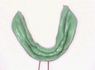
3) An impression taken in Xantopren Green

4) Xantopren impression with beading

5) Addition silicone impression material being used to take an impression of implants

Properties of Addition Silicones

CHEMISTRY

These materials are often termed vinyl polysiloxanes.
Supplied in 2 pastes or in a gun and cartridge form as light, medium, heavy and very heavy bodied.
One paste contains a polydimethylsiloxane polymer in which some methyl groups are replaced by hydrogen. The other paste contains a pre-polymer in which some methyl groups are replaced by vinyl groups, this paste also contains a Chloroplatinic acid catalyst.
On mixing, in equal proportions, crosslinking occurs to form a silicone rubber. Setting occurs in about 6-8 minutes.

PROPERTIES

  • Good shelf life
  • Dimensionally stable
  • Moderate tear strength
  • Excellent surface detail
  • No gas evolution
  • Non toxic and non irritant

ADVANTAGES

  1. Accurate
  2. Ease of use
  3. Fast setting
  4. Wide range of viscosity's

DISADVANTAGES

  1. Hard to mix
  2. Sometimes difficult to remove the impression from the mouth
  3. Too accurate in some circumstances (cast produced is not sufficiently oversized)

Condensation Silicones

CLINICALLY

Used for crown and bridge work mainly, but also for partial dentures, implants and overdentures. Used in stock trays or special trays. One or two stage impression stage. Although dimensionally stable the impression should be cast within 24 hours.

CHEMISTRY

Supplied as a paste and liquid or two pastes, in light, medium, heavy or very heavy bodied (putty).

BASE PASTE

  • Silicone polymer with terminal hydroxy groups
  • Filler


CATALYST PASTE

  • Crosslinking agent (organohydrogen siloxane)
  • Activator (dibutyl-tin dilaurate)

On mixing the two pastes react, cross linking occurs and setting takes about 7 minutes.
The setting reaction is a condensation reaction.
Hydrogen gas is evolved on setting which leads to surface pitting, and a roughened surface to the resulting model.

PROPERTIES

  • Hydrophobic
  • Hydrogen gas evolution on setting
  • Moderate shelf life
  • Moderate tear strength
  • Good surface detail
  • Shrinking of impression over time
  • Non toxic and non irritant
  • Very elastic (near ideal)

ADVANTAGES

  1. Accurate
  2. Ease of use
  3. Can be used on severe undercuts

DISADVANTAGES

  1. Hydrogen evolution
  2. Liquid component of paste/liquid system may cause irritation

Polyethers

Used for crown and bridge work, partial dentures, implants and overdentures. Mixed in a 1:1 ratio until homogeneous colour, the amount of catalyst used can be used to control the setting time. Used in special or stock trays with an adhesive. A one or two stage technique can be used. Although dimensionally stable the impression should be cast within 24 hours.

1) Polyethers come as a two paste system for mixing

Properties of Polyethers

CHEMISTRY

Based on imine chemistry
Supplied in two pastes

BASE PASTE

· Polyether

· Filler

CATALYST PASTE

· Sulphonic acid ester (enhances further polymerisation and crosslinking)

· Inert oils

When mixed the polymer and sulphonic acid ester react to form a stiff polether rubber.
Setting time occurs in about 6 minutes.
Usually only comes in one viscosity - regular bodied, but can also come as light + heavy bodied (Diulent)
Heat and moisture speed up the setting reaction.

PROPERTIES

  • Hydrophillic (ie absorbs water)
  • Good shelf life of up to 2 years
  • Good elastic recovery
  • Non toxic
  • Low setting contraction
  • Low tear strength
  • Excellent surface detail
  • Good dimensional stability

ADVANTAGES

  1. Accuracy
  2. Good on undercuts
  3. Ease of use

DISADVANTAGES

  1. May cause allergic reaction due to the sulphonic acid ester
  2. Poor tear strength
  3. Rapid setting time (ie short working time)
  4. Stiff set material (sometimes hard to remove from mouth)

Elastic Impression Materials-Hydrocolloids-Prosthetic Dentistry Lecture note

Introduction to Elastic Impression Materials
These materials can be stretched and bent to a fairly large degree without suffering any deformation. These are used for recording the patient's mouth where undercuts are present. Usually used for partial dentures, over dentures, implants and crown and bridge work .The elastic impression materials are:

Hydrocolloids

Elastomers

Introduction to Hydrocolloids

A colloid is a state of matter in which individual particles of one substance are uniformly distributed in a dispersion medium of another substance. When the dispersion medium is water it is termed a hydrocolloid. The colloid is relatively fluid when the solute particles present are dispersed throughout the liquid. This is called a sol. Alternatively the particles can become attached to each other, forming a loose network which restricts movement of the solute molecules. The colloid becomes viscous and jelly like, and is called a gel. Some colloids have the ability to change reversibly from the sol state to the gel state. A sol can be converted into a gel in one of two ways:
1. Reduction in temperature, reversible because sol is formed again on heating (eg agar).
2. Chemical reaction which is irreversible (eg alginates). A gel can lose (syneresis which results in shrinkage) or take up (imbibition which results in expansion) water or other fluids.

Hydrocolloids are placed in the mouth in the sol state when it can record sufficient detail, then removed when it has reached the gel state. Hydrocolloid materials especially the alginates, may display a lack of incompatibility with some makes of dental stones. The resultant model may show reduced surface hardness and possibly surface irregularities and roughness.

Agar Impression Materials

CHEMISTRY
  • Agar (colloid)
  • Borax (strengthen gel)
  • Potassium Sulphate
  • Water (dispersion medium)
In its natural state it a gel, but on heating becomes a sol.
PROPERTIES
  • Good surface detail
  • Can be used on undercuts, but liable to tear on deep undercuts
  • Evaporation or imbibition
  • Non toxic and non irritant
  • Slow setting time
  • Poor tear resistance
  • Adequate shelf life
  • Can be sterilised by an aqueous solution of hypochlorite.
ADVANTAGES
1. Good surface detail
2. Reusable and easily sterilised

DISADVANTAGES
1. Need special equipment (water bath) and special technique
2. Dimensional instability

CLINICAL
Supplied in sealed tubes to prevent evaporation of water. The tubes are heated in boiling water (in a water bath) for 10-45 minutes. Once the impression is taken the tray can be cooled with water to aid gel formation. A higher temperature is needed to convert the gel into a sol. The first material to set is that which is in contact with the tray since it is cooler than the tissues. Thus it is the material in contact with the tissue which stays in the sol state for the longest time. Agars have been largely superseded by alginates and elastomers, although are still used for complex impressions for advanced restorative work. They are often used in labs to duplicate model because they can be reused many times.

Alginate Impression Materials

Container of powder should be shaken before use to get an even distribution of constituents. Powder and water should be measured to manufactures instructions. Water at room temperature should be used, this gives a reasonable working time of a couple of minutes. Faster or slower setting times can be achieved by using warm or cold water respectively. The material nearer the tissues sets first (cf. agar). Retention is needed to the impression tray and is provided by perforations in the tray and/or adhesives. Once removed from the mouth the impression should be rinsed with cold water to remove any saliva or blood. It should then be covered in a damp gauze/napkin to prevent syneresis (not placed in water which would cause imbibition-expansion). The impression should be soaked in hypochlorite for 60 seconds and then cast as soon as possible.

1) An alginate impression of the upper arch in a special tray

2) An alginate impression of the lower arch

Properties of Alginates

CHEMISTRY
On mixing the powder with water a sol is formed, a chemical reaction takes place and a gel is formed.
The powder contains

1. Alginate salt (e.g. sodium alginate)
2. Calcium salt (e.g. calcium sulphate)
3. Trisodium phosphate

The setting reaction is as follows:
On mixing the powder with the water
SODIUM ALGINATE

SODIUM SULPHATE
+
>
+
CALCIUM SULPHATE

CALCIUM ALGINATE
The above reaction occurs too quickly often during mixing or loading of the impression tray. It can be slowed down by adding trisodium phosphate to the powder. This reacts with the calcium sulphate to produce calcium phosphate, preventing the calcium sulphate reacting with the sodium alginate to form a gel.
This second reaction occurs in preference to the first reaction until the trisodium phosphate is used up, then the alginate will set as a gel.
There is a well-defined working time during which there is no viscosity change.

PROPERTIES
  • Good surface detail
  • Reaction is faster at higher temperatures
  • Elastic enough to be drawn over the undercuts, but tears over the deep undercuts
  • Not dimensionally stable on storing due to evaporation
  • Non toxic and non irritant
  • Setting time can depend on technique
  • Alginate powder is unstable on storage in presence of moisture or in warm temperatures
ADVANTAGES
1. Non toxic and non irritant
2. Good surface detail
3. Ease of use and mix
4. Cheap and good shelf life
5. Setting time can be controlled with temperature of water used

DISADVANTAGES
1. Poor dimensional stability
2. Incompatibility with some dental stones
3. Setting time very dependent on operator handling
4. Messy to work with


Key Words : dentist find a dentist pediatric dentist cosmetic dentist dentist office family dentist laser dentist sleep dentist sedation dentist painless dentist

Popular Posts

Join This site