Sunday, July 17, 2011

Fundamental Principles of Treatment of Infection-Oral Surgery Lecture Note

In order to treat an acute dent alveolar infection as well as a fascial space abscess correctly, the following are considered absolutely necessary:
·         Take a detailed medical history from the patient.
·         Drainage of pus, when its presence in tissues is established.
This is achieved (1) by way of the root canal, (2) with an intraoral incision, (3) with an extraoral incision, and (4) through the alveolus of the extraction. Without evacuation of pus, that is with administration of antibiotics alone, the infection will not resolve.
·      Drilling of the responsible tooth during the initial phase of inflammation, to drain exudate through the root canal, together with heat therapy. In this way, spread of inflammation is avoided and the patient is relieved of the pain. Drainage may also be performed with trephination of the buccal bone, when the root canal is inaccessible.
·         Antisepsis of the area with an antiseptic solution before the incision.
·     Anesthesia of the area where incision and drainage of the abscess are to be performed, with the block technique together with peripheral infiltration anesthesia at some distance from the inflamed area, in order to avoid the risk of existing microbes spreading into deep tissues.
·         Planning of the incision so that:
– Injury of ducts (Wharton, Stensen) and large vessels and nerves is avoided.
– Sufficient drainage is allowed. The incision is performed superficially, at the lowest point of the accumulation, to avoid pain and facilitate evacuation of pus under gravity.
– The incision is not performed in areas that are noticeable, for esthetic reasons; if possible, it is performed intraorally.
·       Incision and drainage of the abscess should be performed at the appropriate time. This is when the pus has accumulated in the soft tissues and fluctuates during palpation, that is when pressed between the thumb and middle finger, there is a wave-like
Incision for drainage of a sublingual abscess. The incision is performed parallel to the submandibular duct and the lingual nerve

Incision for drainage of a palatal abscess, parallel to the greater palatine vessels
Incisions for drainage of a submandibular or parotid (a), and a submasseteric (b) abscess. During cutaneous incisions, the course of the facial artery and vein must be taken into consideration (a), as well as that of the facial nerve (b)
movement of the fluid inside the abscess. If the incision is premature, there is usually a small amountof bleeding, no pain relief for the patient and the edema does not subside.
·       The exact localization of pus in the soft tissues (if there is no fluctuation present) and the incision for drainage must be performed after interpretation of certain data; for example, ascertaining the softest point of swelling during palpation, redness of the skin or mucosa, and the most painful point to pressure. This area indicates where the superficial incision with a scalpel is to be made. If there is no indication of accumulation of pus to begin with, hot intraoral rinses with chamomile are recommended to speed up development of the abscess and toensure that the abscess is mature.
·       Avoid the application of hot compresses extraorally, because this entails an increased risk of evacuation of pus towards the skin (spontaneous drainage)
Superficial incisions on the skin (a) and on the mucosa of the oral cavity (b)
Spontaneous extraoral (undesirable) drainage of an abscess, after the erroneous placement of hot compresses on the skin
·     Drainage of the abscess is initially performed with a hemostat, which, inserted into the cavity of the abscess with closed beaks, is used to gently explore the cavity with open beaks and is withdrawn again with open beaks. At the same time as the blunt dissection is being performed, the soft tissues of the region are gentlymassaged, to facilitate evacuation of pus.
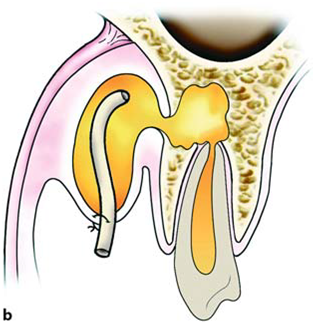
·      Placement of a rubber drain inside the cavity and stabilization with a suture on one lip of the incision, aiming to keep the incision open for continuous drainage of newly accumulated pus.
·        Removal of the responsible tooth as soon as possible, to ensure immediate drainage of the inflammatory material, and elimination of the site of infection. Extraction is avoided if the tooth can be preserved, or if there is an increased risk of serious complications in cases where removal of the tooth is extremely difficult.
·   Administration of antibiotics, when swelling is generally diffuse and spreading, and especially if there is fever present, and infection spreads to the fascial spaces, regardless of whether there is an indication of the presence of pus. Antibiotic therapy is usually empiric, given the fact that it takes time to obtain the results from a culture sample. Because the microorganisms isolatedmost often in odontogenic infections are streptococci (aerobic and anaerobic), penicillin remains the antibiotic of choice for treatment.
Diagrammatic illustrations showing the incision of an intraoral abscess and the placement of a hemostat to facilitate the drainage of pus
Diagrammatic illustrations showing the placement of a rubber drain in the cavity and stabilization with a suture on one lip of the incision.
Treatment of Infection in Cellular Stage
In this stage, treatment of the infection depends on the location of existing pus. Localization, as already mentioned, may be intraalveolar, subperiosteal, submucosal or subcutaneous. Each of these cases is discussed in next posts.

Key Words : oral surgeons wisdom teeth extraction dental implant surgery doctor of dental surgery oral surgeon dental operation dental gum surgery wisdom tooth surgery dental surgery oral surgery doctor of dental surgery cosmetic dental surgery dental laser surgery cosmetic gum surgery laser gum surgery

Saturday, July 16, 2011

Immediate Denture-Prosthetic Dentistry Lecture note and Power Point Presentation(PPT) Free Download


Immediate denture
All the dentures that you made in this year is a conventional dentures for edentulous patients which means they have been extracted their teeth then they seek to fabricate a dentures
Now we deal with patients whose don’t want to extract their teeth before fabricate the denture( he can't go out and continue to exercise their life without teeth) so in this case you fabricate the denture first then teeth extraction and denture insertion done at the same day

Immediate denture :
* is complete denture or removable partial denture fabricate for replacement immediately after the removal of natural teeth
*it is a denture constructed before the extraction of the teeth which it replaces and fitted immediately after the teeth extraction

Keep in your mind that in immediate denture you will deal with dentate patient means you will fabricate denture for dentate patient not for edentulous patient and teeth extraction will be at the delivery stage and it can be complete or partial

again the patient come to you and he still has his teeth , the teeth need to be extracted but the patient refuse to life as edentulous , he want to receive his denture immediately after the extraction but as you do and as you know , denture needs from 5 to 6 visits to be finished so what can
you do??? it is immediate denture means you will not extract the teeth , you just take an impression inorder to fabricate denture for the patient as what we do in partial denture then at the time of delivery you will extract the teeth and patient get his denture and go home with teeth and don’t life as edentulous .sooooooo
denture making => teeth extraction => denture insertion

Type of immediate denture
*transitional /interim
*conventional
*post immediate

transitional /interim; you remember in RPD there is one type of it which called transitional partial denture and the reason why we fabricate it because there are some of teeth will going to be extracted sooooooo once you diced to extract the teeth ,the transitional partial denture have to be converted to complete denture which called transitional immediate complete denture and as we said its immediate means at the same time where you extract the teeth patient will get his denture

conventional immediate complete denture; here the patient come to extract his teeth (don’t have RPD)but he want to receive his denture immediately after extraction soooo what you will do for him is conventional immediate denture
usually when we say immediate complete denture we mean the conventional type

now how to make immediate denture

Technique
· Patient examination and diagnosis then put your treatment plane
· Informed consent ; means you have to explain for your patient what the advantages and disadvantages of immediate denture ,it is more expensive ,the denture may need to rebase in a few month and it is necessary to replace the denture at a later period
· Oral hygiene procedure ; the teeth should scaled and cleaned and oral hygiene brought up
· Extract some posterior teeth so you need to decide which teeth need to be extracted and which teeth retained as guide to correct jaw relation , regarding to the anterior teeth keep the incisor and canine without extraction to coincide with stable intercuspal occlusion
Note; the extraction of posterior teeth done through more than two visits , one side at each visit and interdentally and interradicular bone should be smoothen after the extraction .this unilateral extraction and care of each part promote healing and reduce the time of partially edentulous and give me a better support
· Wait 4-8 weeks for healing
· Take prelimary impression and as you know you take an impression for patient with teeth ( remember anterior teeth still there and some posterior teeth )so we use an elastic material which is alginate or rubber base and use stock tray for dentate patient (don’t forget that you deal with dentate patient ) then we send it to the lab to fabricate special tray with spacer (we need spacer for alginate impression that will use in final impression )
Note;
1. in immediate denture we totally exclude ZO eugenol material cause you deal with dentate patient and the best impression taken by alginate
2. if the patient has transitional partial denture we take the impression while he wear his denture , the same thing in the final impression
3. in post immediate denture , patient may have bridge or implants so we take the impression over it
· Take the final impression by alginate or elastomer or we can use alternative impression which is a compression impression , it composition of impression past (compound impression) for edentulous part and alginate for dentate part which means , take the impression by compound for edentulous part then put alginate over the compound and retake the impression then we send it to the lab to make record block
· Jaw relation record then mount the casts together and start to set up the teeth (the only missing teeth is set and the teeth that are not extracted we don’t set it ) and go for tray-in visit but here in immediate denture , not all the teeth is set so the tray- in is consider partially not completely tray-in cause I can't check the aesthetic and phonetics ,that’s why we consider tray-in visit is lost in immediate complete denture ,actually we do this visit but we miss some steps as aesthetic and phonetics because the anterior teeth still natural , it is not a part of the denture and in this visit I just check the teeth that is stetted in the edentulous area which is the posterior teeth
Now I finish tray- in and determine the post dam area we go to insertion visit and as we said we insert the denture and extract the teeth at the same visit sooo until now the denture not complete it still partial and I want to get it for the patient complete but how????
Again until now I do all the steps for the patient and the next visit is the extraction and insertion but the denture is still partial it is not complete and next visit the patient will get his complete denture not partial, so how to convert the denture into complete ????
We ask the technician to remove the remaining teeth from master cast which is usually canines and incisors and then add teeth and acrylic to the existing denture but to remove teeth from the cast I need reference point means I need to tell the technician how much he cut from the tooth and how much he go in the dental pocket!!!!!
Assume the patient has periodontal problem he will has pocket depth more than healthy patient so you need to tell your technician how much he go deep in dental pocket
Another thing you need to think about it is the design of immediate denture cause patient will wear the denture at the same time of extraction and the main problem here is maxillary prominence ,so we need to think how to fit the denture in this prominent premaxilla ,we have 4 options,
1. Open face design (without anterior flange) it give me natural appearance and it easy to insert , exactly reproduction of the teeth position and easier to set the teeth and no interference with lip musculature but it give me poor retention , the natural appearance is not long maintained ,short life denture , difficult to rebase and irregularity of anterior ridge may develop
2. Labial flange without alveolectomy ; good retention and support ,easy to rebase , give me strong denture and rapid healing with smooth ridges but it difficult on case of undercut and poor appearance
3. Labial flange with alveolectomy; it indicate in case of
1-prominent pre-maxilla which prevent denture insertion and give me poor appearance
2-limited anterior interalveolar space and deep vertical overlap
4. Labial flange with alveoletomy ; we use it if the alveolectomy is carried out and we need to eliminate the undercut but sometimes the undercut can't be completely eliminated
Alveolectomy is totally remove of labial plate( you will remove from the cortical bone )
Alveoletomy just remove the interseptal bone(the bone that remain between the sockets of the teeth after extraction ) and collapse the labial plate into the palatal plate so labial plate will pushed backward so you cut from sponge bone
So you need to tell the technician which design you select and he will adjust it on the cast how??
If you decide to do alvectomy the technician should trim all labial plate from the cast or if you decide to do alvelotomy he will trim less from the cast and if you don’t want to any of them just tell the technician how much he cut from the socket and he will adjust it
again you need to tell your technician how much he cut from the teeth and how much he go deep on the packet and which design you decide to do and he will adjust all of these on the cast .
After he cut the teeth from the cast and adjust the design and the deep of socket , start to set the teeth and do all the processing ,packing and fasking
Now the denture is ready to insert in patient mouth and we go to the next visit (extraction and insertion )
· Extraction and insertion
we send patient to the surgeon to extract his remaining teeth resulting in residual ridge but did you think the residual ridge that remain after teeth extract is the same ridge that the technician was designed on the cast as you tell him ?????????? really it not because sometimes extraction leave bone spicules or high spot in some area so who we can make the ridge of the patient similar to ridge in the cast ?????
After the technician cut the teeth from the cast and adjust the design he should take impression for the cast to acts as a surgical guide by acrylic or silicon , so after teeth extraction, the surgeon will take this guide and adapt it on the ridge and trim from the bone or from the tissues where there is pressure , this way will help us specially if the design is alveolctomy cause it more likely to leave spicules
So by this way the ridge have been adjusted in the clinic exactly as has been adjusted in the lab on the cast
· Post insertion care
After denture inserted to patient mouth you have to instruct your patient to keep the denture inside his mouth for 24 hours and come next day without remove it and tell him if it loose-out suddenly during this time put it back immediately because there is injury in the tissue caused by the extraction and remove the denture may cause edema in addition to avoid rising first day
So the patient will came back after 24 hours and usually there is high spot (red spot ) we relive it , don’t use pressure indicating past cause it may causing pressure over the healing socket , alternative of it we use indelible pencil
After that patient can get his denture off and start to rise his mouth by water and salt or to clean the denture and sleep while get it off
3-5 days ,bring your patient again , use pressure indicting past and do slight refine the occlusion
Follow up, you can use tissue condition if need and remove any socket convexities to avoid healing defect
You may notice that the denture become loose , this is because the patient used his denture so there will be some changes on the alveolar ridge ( as you know bone resorption following extraction of the teeth is rapid at first ,but it continues at a reduced rate throughout life )and this will cause loss in retention , to improve that we use tissue conditioner
And keep in your mind immediate denture need to be reline and rebase 3 months later on (only necessary in the area of extraction) and replace by a new denture during 6 to 12 months(6-12 months is the time of the residual ridges resorption and after that the denture become loss and should to replace it)

Post immediate denture
rarely used , Its idea is the patient don’t want to extract his posterior teeth( usually he has extensive bridge from 3 to 7 or implant or there is pathology such as a large cyst ) means he want to extract all his teeth in the delivery stage and this will eliminate tray- in stage so you need to be more accurate and there is a high chance of error
after we show the types and technique of immediate denture what is the advantages and disadvantages of it
Advantages
* self esteem => one of the main problem with complete denture is the patient become disappointment once they loss their teeth and they can't socialized and eating or speaking but when the denture is immediate the edentulous period is eliminated and this has a great social and psychological significant
* jaw relation are kept => cause the patient still has his teeth so you can check centric relation and vertical diminition perfectly
* function is maintained => patient keep eating and speaking and do all the function until the insert visit and after that he will start using his denture so he will not leave your clinic with any gap or any tooth loss during the procedure
* aesthetic => cause he never life as a edentulous
* less resorption with rapid healing => as you know as long as patient become edentulous bone become more resorp and this is caused by the load on alveolar ridge but when the patient get denture immediately after teeth extraction the resorp will be less

Notes
Bone resorption is happen due to the load over the ridge which stimulate bone resorp and this load is less if the patient wear his denture immediately
* a very natural and functional result can be obtained as lip position .occlusal plane , vertical height and occlusion can all be exactly
* functional mastication and tone of facial muscles is maintained
* size .shape ,shade and position of teeth can be accurately reproduced
*no unnatural mandibular movement will developed and change in tongue shape is prevented
*little interference with speech , diet ,TMJ and its function
*socket are protected and healing is quickened

Disadvantages
*if there is slight malocclusion the denture become unstable means if natural teeth are maloccluded then the accurate reproduction is not possible so the result is prosthetic malocclusion and irregular occlusal plane
*it need more time and more appointment in addition it more cost
* there is no try- in stage ; as we said previously that actually you do tray-in visit but it lost some steps as aesthetic and phonetics so you need to be more accurate in your work
*can't be made for all type of patient some of them is irresponsible and wear immediate denture for long time will damage the oral cavity
 
Free Download Immediate Denture
PowerPoint Presentation(PPT) Here


Tags : Immediate denture,Prosthetic dentistry lectures notes,Prosthodontic lecture notes,Denture design lectures

Friday, July 15, 2011

Odontogenic Infections-Oral Surgery Lecture Note

In order to understand how odontogenic infections are treated, the dentist must be familiar with the terminology concerning infection and the pathophysiology of inflammation, which are described below.
Inoculation
is characterized by the entry of pathogenic microbes into the body without disease occurring.An infection involves the proliferation of microbes resulting in triggering of the defense mechanism, a process manifesting as inflammation.
Inflammation
is the localized reaction of vascularand connective tissue of the body to an irritant, resulting in the development of an exudate rich in proteins and cells. This reaction is protective and aims at limiting or eliminating the irritant with various procedures while the mechanism of tissue repair is triggered. Depending on the duration and severity, inflammation is distinguished as acute, subacute or chronic.
Acute Inflammation
This is characterized by rapid progression and is associated with typical signs and symptoms. If it does not regress completely, it may become subacute or chronic.
Subacute Inflammation
This is considered a transition phase between acute and chronic inflammation.
Chronic Inflammation
This procedure presents a prolonged time frame with slight clinical symptoms and is characterized mainly by the development of connective tissue. Inflammation may be caused by, among other things, microbes, physical and chemical factors, heat, and irradiation. Regardless of the type of irritant and the location of the defect, the manifestation of inflammation is typical and is characterized by the following clinical signs and symptoms: rubor (redness), calor (heat), tumor (swelling or edema), dolor (pain), and functio laesa (loss of function).
The natural progression of inflammation is distinguished into various phases. Initially vascular reactions
with exudate are observed (serous phase), and then the cellular factors are triggered (exudative or cellular phase). The inflammation finally resolves and the destroyed tissues are repaired. On the other hand, chronic inflammation is characterized by factors of reparation and healing. Therefore, while acute inflammation is exudative, chronic inflammation is productive (exudative and reparative). Understanding the differences between these types of inflammation is important for therapeutic treatment. Serous Phase. This is a procedure that lasts approximately 36 h, and is characterized by local inflammatory edema, hyperemia or redness with elevated temperature, and pain. Serous exudate is observed at this stage, which contains proteins and rarely polymorphonuclear leukocytes.
Cellular Phase.
This is the progression of the serous phase. It is characterized by massive accumulation of polymorphonuclear leukocytes, especially neutrophil granulocytes, leading to pus formation. If pus forms in a newly developed cavity, it is called an abscess. If it develops in a cavity that already exists, e.g., the maxillary sinus, it is called an empyema.
Reparative Phase
During inflammation, the reparative phenomena begin almost immediately after inoculation. With the reparative mechanism of inflammation, the products of the acute inflammatory reaction are removed and reparation of the destroyed tissues follows. Repair is achieved with development of granulation tissue, which is converted to fibrous connective tissue, whose development ensures the return of the region to normal.
Infections of the Orofacial Region
The majority (i.e., 90–95%) of infections that manifest in the orofacial region are odontogenic. Of these, approximately 70% present as periapical inflammation, principally the acute dentoalveolar abscess, with the periodontal abscess following, etc.
Periodontal abscess originating from a maxillary central incisor.
Radiograph of same case showing bone resorption, which led to the formation of a periodontal pocket
Etiology
The cardinal causes of orofacial infections are non-vital teeth, pericoronitis (due to a semi-impacted mandibular tooth), tooth extractions, periapical granulomas that cannot be treated, and infected cysts. Rarer causes include postoperative trauma, defects due to fracture, salivary gland or lymph node lesions, and infection as a result of local anesthesia.
Periodontal Abscess
This is an acute or chronic purulent inflammation, which develops in an existing periodontal pocket. Clinically, it is characterized by edema located at the middle of the tooth, pain, and redness of the gingiva. These symptoms are not as severe as those observed in the acute dentoalveolar abscess, which is described below.
Treatment of the periodontal abscess is usually simple and entails incision, through the gingival sulcus with a probe or scalpel, of the periodontal pocket that has become obstructed. Incision may also be performed at the gingivae; more specifically, at the most bulging point of the swelling or where fluctuation is greatest.
Acute Dentoalveolar Abscess
This is an acute purulent inflammation of the periapical tissues, presenting at nonvital teeth, especially when microbes exit the infected root canals into periapical tissues. Clinically, it is characterized by symptoms that are classified as local and systemic.
Local Symptoms
Pain
The severity of the pain depends on the stage of development of the inflammation. In the initial phase the pain is dull and continuous and worsens during percussion of the responsible tooth or when it comes into contact with antagonist teeth. If the pain is very severe and pulsates, it means that the accumulation of pus is still within the bone or underneath the periosteum. Relief of pain begins as soon as the pus perforates the periosteum and exits into the soft tissues.
Edema
Edema appears intraorally or extraorally and it usually has a buccal localization and more rarely palatal or lingual. In the initial phase soft swelling of the soft tissues of the affected side is observed, due to the reflex neuroregulating reaction of the tissues, especially of the periosteum. This swelling presents before suppuration, particularly in areas with loose tissue, such as the sublingual region, lips, or eyelids. Usually the edema is soft with redness of the skin. During the final stages, the swelling fluctuates, especially at the mucosa of the oral cavity. This stage is considered the most suitable for incision and drainage of the abscess.
Other Symptoms
There is a sense of elongation of the responsible tooth and slight mobility; the tooth feels extremely sensitive to touch, while difficulty in swallowing is also observed.
Systemic Symptoms
The systemic symptoms usually observed are: fever, which may rise to 39–40 °C, chills,malaise with pain inmuscles and joints, anorexia, insomnia, nausea, and vomiting. The laboratory tests show leukocytosis or rarely leukopenia, an increased erythrocyte sedimentation rate, and a raised C-reactive protein (CRP) level.
Complications
If the inflammation is not treated promptly, the following complications may occur: trismus, lymphadenitis at the respective lymph nodes, osteomyelitis, bacteremia, and septicemia.
Diagnosis
Diagnosis is usually based upon clinical examination and the patient’s history. What mainly matters, especially in the initial stages, is the localization of the responsible tooth. In the initial phase of inflammation, there is soft swelling of the soft tissues. The tooth is also sensitive during palpation of the apical area and during percussion with an instrument, while the tooth is hypermobile and there is a sense of elongation. In more advanced stages, the pain is exceptionally severe, even after the slightest contact with the tooth surface. Tooth reaction during a test with an electric vitalometer is negative; however, sometimes it appears positive,which is due to conductivity of the fluid inside the root canal.
Radiographically, in the acute phase, no signs are observed at the bone (whichmay beobserved 8–10 days later), unless there is recurrence of a chronic abscess,where upon osteolysis is observed. Radiographic verification of a deeply carious tooth or restoration very close to the pulp, as well as thickening of the periodontal ligament, are data that indicate the causative tooth.
Differential diagnosis of the acute dentoalveolar abscess includes the periodontal abscess, and the dentist must be certain of his or her diagnosis, because treatment between the two differs.
Spread of Pus Inside
Tissues From the site of the initial lesion, inflammation may spread in three ways:
1. By continuity through tissue spaces and planes.
2. By way of the lymphatic system.
3. By way of blood circulation.
Diagrammatic illustrations showing spread of infection (propagation of pus) of an acute dentoalveolar abscess,depending on the position of the apex of the responsible tooth. a Buccal root: buccal direction.b Palatal root: palatal direction
The most common route of spread of inflammation is by continuity through tissue spaces and planes and usually occurs as described below. First of all, pus is formed in the cancellous bone, and spreads in various directions by way of the tissues presenting the least resistance.
a Spread of pus towards the maxillary sinus, due to the closeness of the apices to the floor of the antrum.b Diagrammatic illustration showing the localization of infection above or below the mylohyoid muscle, depending on the position of the apices of the responsible tooth
Whether the pus spreads buccally, palatally or lingually depends mainly on the position of the tooth in the dental arch, the thickness of the bone, and the distance it must travel.Purulent inflammation that is associated with apices near the buccal or labial alveolar bone usually spreads buccally, while that associated with apices near the palatal or lingual alveolar bone spreads palatally or lingually respectively. For example,the palatal roots of the posterior teeth and the maxillary lateral incisor are considered responsible for the palatal spread of pus, while the mandibular third molar and sometimes the mandibular second molar are considered responsible for the lingual spread of infection. Inflammation may even spread into the maxillary sinus when the apices of posterior teeth are found inside or close to the floor of the antrum. The length of the root and the relationship between the apex and the proximal and distal attachments of various muscles also play a significant role in the spread of pus. Depending on these relationships, in the mandible pus originates from the apices found above themylohyoid muscle, and usually spreads intraorally, mainly towards the floor of the mouth (sublingual space). When the apices are found beneath the mylohyoid muscle (second and third molar), the pus spreads towards the submandibular space, resulting in extraoral localization. Infection originating from incisors and canines of the mandible spreads buccally or lingually, due to the thin alveolar bone of the area. It is usually localized buccally if the apices are found above the attachment of the mentalis muscle. Sometimes, though, the pus spreads extraorally, when the apices are found beneath the attachment. In the maxilla, the attachment of the buccinators muscle is significant.When the apices of the maxillary premolars and molars are found beneath the attachment of the buccinator muscle, the pus spreads intraorally; however, if the apices are found above its attachment, infection spreads upwards and extraorally. Exactly the same phenomenon is observed in the mandible as in the maxilla if the apices are found above or below the attachment of the buccinator muscle.
Spread of pus depending on the length of root and attachment of buccinators muscle. a Apex above attachment: accumulation of pus in the buccal space. b Apex beneath the buccinator muscle: intraoral pathway towards the mucobuccal fold
In the cellular stage, depending on the pathway and inoculation site of the pus, the acute dentoalveolar abscessmay have various clinical presentations, such as:(1) intraalveolar, (2) subperiosteal, (3) submucosal, (4) subcutaneous, and (5) fascial ormigratory – cervicofacial.

Intraalveolar abscess of maxilla (a) and mandible (b). Diagrammatic illustrations show accumulation of pus at a portion of the alveolar bone in relation to the periapical region
The initial stage of the cellular phase is characterized by accumulation of pus in the alveolar bone and is termed an intraalveolar abscess . The pus spreads outwards from this site and, after perforating the bone, spreads to the subperiosteal space, from which the subperiosteal abscess originates, where a limited amount of pus accumulates between the bone and periosteum. After perforation of theperiosteum, the pus continues to spread through the soft tissues in various directions. It usually spreads intraorally, spreading underneath the mucosa forming the submucosal abscess.
Subperiosteal abscess with lingual localization. a Diagrammatic illustration; b clinical photograph
Sometimes, though,it spreads through the loose connective tissue and, after its pathway underneath the skin, forms a subcutaneous abscess, while other times it spreads towards the fascial spaces, forming serious abscesses called fascial space abscesses.
Subcutaneous abscess originating from a mandibular tooth. a Diagrammatic illustration. b Clinical photograph.The swelling mainly involves the region of the angle of the mandible
The fascial spaces are bounded by the fascia, which may stretch or be perforated by the purulent exudate, facilitating the spread of infection. These spaces are potential areas and do not exist in healthy individuals, developing only in cases of spread of infection that have not been treated promptly. Some of these spaces contain loose connective tissue, fatty tissue, and salivary glands,while others contain neurovascular structures. Acute diffuse infection,which spreads into the loose connective tissue to agreat extent underneath the skin with or without suppuration,is termed “cellulitis” (phlegmon), and is described in next posts.
Fascial abscess (submandibular). a Diagrammatic illustration. b Clinical photograph
Next Post : Fundamental principles of treatment of infection
Tags : Odontogenic infections, Infections of Oromaxillofacial Region, Infection from lower 3rd molar,Spread of infections,Factors determining spread of infections,Spread of Infections.

Fundamental Principles of Treatment of Infection-Oral Surgery Lecture 

Key Words : oral surgeons wisdom teeth extraction dental implant surgery doctor of dental surgery oral surgeon dental operation dental gum surgery wisdom tooth surgery dental surgery oral surgery doctor of dental surgery cosmetic dental surgery dental laser surgery cosmetic gum surgery laser gum surgery


Popular Posts

Join This site