Thursday, June 7, 2012

Concept of Neutral Zone

Definition
‘The neutral zone is that area in the potential denture space where the forces of tongue pressing outward are neutralized by the forces of the cheeks and lips pressing inward’.
Since these forces are developed through muscular contraction during the various functions of chewing, speaking, and swallowing, they vary in magnitude and direction in different individuals.


The Potential ‘Denture Space’
The central thesis of the neutral zone approach to complete dentures is ‘to locate that area in the edentulous mouth where the teeth should be positioned so that the forces exerted by the muscles will tend to stabilize the denture rather than unseat it’.
The soft tissues that form the internal and external boundaries of the denture space exert forces which generally influence the stability of the dentures.

Importance of Neutral Zone
During childhood, the teeth erupt under the influence of muscular environment created by forces exerted by tongue, cheeks and lips, in addition to the genetic factor. These forces has a definite influence upon the position of the erupted teeth, the resultant arch form, and the occlusion.
Generally, muscular activity and habits which develop during childhood continue through life and after the loss of teeth, it is important that the artificial teeth be placed in the arch form compatible with these muscular forces.

As the area of the impression surface decreases (due to alveolar ridge resorption), less influence it has on the denture retention and stability.

Consequently, retention and stability become more dependent on the correct positioning of the teeth and the contours of the external or polished surfaces of the dentures.
Therefore, these surfaces should be so contoured that the horizontally directed forces applied by the peri -denture muscles should act to seat the denture.

The Neutral-Zone Philosophy

is based upon the concept that for each individual, there exists within the denture space a specific area where the function of the musculature will not unseat the denture & where forces generated by the tongue are neutralized by the forces generated by lips and cheeks.
The artificial teeth should not be placed on the crest of the ridge or buccally or lingually to it – rather these should be placed as dictated by the musculature.
The objectives achieved by this approach are,
                a) the teeth will not interfere with the normal muscle function, &
                b) the forces generated by these muscles against the denture, especially for the resorbed lower ridge, are more favorable for stability & retention.

Muscles involved in the ‘Neutral Zone’
The musculature of the denture space can be divided into two groups,
                1. those muscles which primarily dislocate the denture during activity (Dislocating muscles),
                2. those muscles that fix the denture by muscular pressure on the polished surfaces (Fixing muscles).
These can then be further divided according to their location on the vestibular (labial & buccal) side or lingual side of the dentures.

Dislocating muscles
                Vestibular:
                                Masseter
                                Mentalis
                                Incisive Labii Infer.
                Lingual:
                                Medial Pterygoid
                                Palatoglossus
                                Styloglossus
                                Mylohyoid
Fixing muscles
                Vestibular:
                                Buccinator
                                Orbicularis oris
                Lingual:
                                Genioglossus
                                Lingual longitudinal
                                Lingual vertical
                                Lingual transverse

Technique for  the  Location  of  Neutral  Zone
A number of variations of the basic technique have been reported in the literature. However, with all these techniques of neutral zone approach, the usual sequence of complete denture construction is somewhat reversed.
1. Individual trays are constructed and adjusted carefully in the mouth so that these are stable on opening the mouth, speaking, and swallowing.
2. Modeling compound is used to fabricate occlusion rims.
3. These rims are then molded intra orally according to the muscle function – recording of neutral zone.
4.  Establishing the tentative OVD and CR.
5.  Obtain the final impression with the closed mouth technique.
6.  Final determination of the OVD and CR.
7.  Pouring the casts, forming the plaster index, their articulation, and Set-up of the teeth.
8.  Wax try-in of the dentures and verification of the tooth position intra-orally.
9.  Finally, obtaining the impression of the polished surfaces and establishing their contours in the wax-up.
Recording the Neutral Zone
 

Jaw relation records & reference lines
Plaster index fabrication and tooth arrangement

Tooth arrangement & initial wax-up for the soft tissue contours – lingual index removed
Tooth arrangement in the Neutral Zone Buccal Plaster indices are being removed

Waxed complete dentures Intra oral Try – in
Recording Neutral Zone - Soft tissue Contours
Application of Vaseline before adding impression material  
Impression material is evenly applied on the buccal and lingual surfaces of the waxed-up dentures

Patient performs oral functions including chewing to determine the thickness, contour and shape of the polished surfaces

Carefully inspect the impression of the polished surfaces including the palatal area – for complete coverage by the impression material

The material flown over the tooth surfaces must be removed carefully with a carver

The Finished Complete Dentures based on the Neutral Zone Concept


Recording Neutral Zone for a Single Complete Denture
Occlusal stops established intra-orally and retentive wire added to the special tray
Slow setting medium viscosity silicone impression material is coated on all the surfaces.
After inserting the tray, patient is advised to smile, swallow and to produce vowels, ‘ooh, ah’, until the material is set.

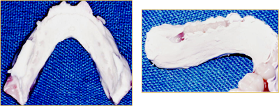
Denture space Impression after removal from the mouth

Its appearance is totally un-conventional. Any evidence of large areas of air entrapment, insufficient or excessive volume of impression material, or tray showing through necessitate re-taking the impression.
The Poured Denture space Impression-Four matrices are required to record the buccal, labial, lingual & ridge contours

The impression seated on the ridge matrix (with the buccal, labial and lingual matrices removed) is mounted against the upper cast on the articulator.

Silicone impression is then removed – buccal and labial matrices (surfaces) are replaced. 
Softened wax is then placed in the space for setting the lower teeth for wax try- in.

The Waxed Trial Denture

The soft tissue contours are carefully developed without altering the basic contours of the recorded impression.
The routine assessments are conducted at the trial insertion, with special emphasis on the stability of the denture.

Some other techniques for recording Neutral Zone
Different designs of Impression trays

Injecting the Alginate into the Denture space ‘Impression tray is stabilized by biting’

Articulation & Set-up of teeth

Alginate impression acts as the index for tooth position
Replacing Impression material with Wax rim Setting the teeth with a plaster index


Further Applications of the Basic Technique

Determining the Fit of a completed denture to the Neutral Zone
Coat the polished surfaces of the denture with a low viscosity silicone impression material. Ask the patient to perform functional movements while the material sets. Inspect the denture & adjust any heavy muscle contact.


Determining the optimal space for a segment of the denture
Remove the teeth and the base material from the segment of the denture that needs modification. Apply adhesive and take the impression with moldable material. Check for stability and undertake the laboratory procedures.
Neutral Zone Versus Biometrics
Neutral Zone concept for the placement of artificial teeth could not enjoy the universal approval as did the Biometric concept of tooth arrangement. The reasons for this limited success are numerous, e.g.,
1. The viscosity of the material used for obtaining this impression is critical. More viscous the material, more it will be difficult for the muscles to mold it and visa versa.
2. The geriatric patients could suffer difficulty during the procedure as their musculature may have lost the tone.
3. The stability and retention of the bases on the soft denture support must be excellent as well as the comfort.
4. The resultant ‘neutral zone’ is often narrow and more lingually placed - with the closed mouth technique, the tongue could not perform all the functional movements, such as the phonetics.
5. This technique does not offer any guidelines for the selection of the teeth.
6. The technique is troublesome for the patient and does not offer much advantage over the biometric guides for the arrangement of teeth.

Wednesday, June 6, 2012

Clinical stages in Removable partial denture construction


This guide will assist you in clinical stages of removable partial denture construction. You will, benefit by following the approaches suggested in this document.   It must be  understood  however,  that  there  may  be  necessary  modification  required  through  situations which  are  without  your  control.   Such  modification  should  always  be  discussed  with  and  agreed by your clinical teacher.

1st visit
Preliminary Impressions 

   Selection of stock tray. Modification  of  the  tray  with  impression  compound  or autopolymerising  acrylic  as appropriate.
     Normally a high  viscosity alginate should be used as this will compensate for  the lack of  fit  of  the  stock  tray.   A  thin  layer  of  adhesive  should  be  applied  to  the  tray  before starting to mix the alginate. 
Prescription

It  is  essential  at  this  and  subsequent  stages  to  indicate  precisely  what  is  required  for  the  next  appointment. The prescription on the laboratory card must be clear and comprehensive. If there is
any  possibility  of  confusion  it  is    essential  to  discuss  the  case  personally  with  the  technician  involved.   All  casts  at  this  stage  should  be  poured  in  dental  stone and the type and material of  individual trays indicated.

If the laboratory card is not completed and dated, work will not be available for the next  appointment.

Design

The  design  of  a  partial  denture  should  be  determined  before  master  impressions are recorded.In this respect casts where there are opposing natural teeth in contact, casts must be mounted on
an  articulator  and  surveyed  to  produce  the  desired  design.      In  many  cases  where  there  are  sufficient teeth, casts can be placed in occlusion by hand prior to mounting.In other situations it  will be unnecessary to construct occlusal rims to register the jaw relationship of the patient. 


The sequence therefore is as follows:-

     Pour preliminary casts.
     Survey initially to vertical path of insertion.
     In very few cases a second survey will be necessary to a modified path of insertion.
     Mount casts on articulator.
     Produce design for removable partial denture.

The  provisional  design  produced  should  be  discussed  with  your  clinical  teacher  before  the patient's  next  appointment.   At  this  stage  a  decision  should  be  made on  the  need  for  possible  tooth preparation or modification.  This may indicate that the following may be necessary:-

•     Rest  seat  preparation    to  provide  sufficient  space  and  a  horizontal  surface  for  any  support component.
•     Modification of tooth contour, by grinding or the addition of light-cured composite resin, to improve the action of clasp arms or the occlusal relationship.

The  proposed  design  should   be  drawn  on  the  laboratory  card  and also transferred to the study cast which should be retained for reference until the trial stage has been completed.   The design prescription must be clear and comprehensive.

If  surgical,  conservative  or  periodontal    treatment  is  indicated  this  must  be  completed    before recording  master  impressions   This  is  because  the  type  of  denture  required  may  influence  the overall  treatment  plan,  e.g.  rest  seats  incorporated  into  Class  II  restorations,  full  veneer  crowns contoured to provide undercut areas for retention, or tooth extraction as a result of over eruption.

2nd Visit
Normally the second visit will be for master impressions if the preliminary casts have already been mounted and a design determined.   In cases where the preliminary casts could not be mounted however,  the  second  visit  will  be  devoted  to  recording the jaw relationship of the patient prior to mounting casts on the articulator and developing a design.

Recording Jaw Relationships

If an occlusal stop is present in the mouth you must determine whether the associated intercuspal position is acceptable.   If there is horizontal (antero-posterior or lateral) deviation of the mandible after the initial occlusal contact, it may be necessary to correct the deflective occlusal contact by tooth   modification,   extraction   or   (rarely)   orthodontic   treatment.     If   there   is   loss   of   vertical dimension of   occlusion (OVD) the appropriate increase will have to be determined by adjusting
occlusal rims in relation to the rest  vertical dimension (RVD).


For the purpose of jaw relationships and their registration partially dentate patients can be divided into two categories:-
·         Patients without an occlusal stop to indicate the correct intercuspal position or vertical dimension of occlusion.
·         Patients with occlusal contact in the intercuspal position. 


First Category

In the first category the OVD is determined by establishing   the RVD and modifying the occlusal rims  until  the  OVD  is    some  2-4mm  short  of  the  RVD,  this  distance  indicating  the  amount  of interocclusal clearance.   The horizontal jaw relationship recorded should be the retruded contact position.

  • Occlusal rims (and wax trial dentures) should only be placed in the mouth long enough to carry out a particular clinical procedure.    On removal they should be chilled in a bowl of cold water to avoid distortion 
  • In  the  mouth  the  fit  and  extension  of  the  rim  should  be  checked  and  modified  if necessary to produce acceptable stability. 
  • The  upper  occlusal  rim  should  be  adjusted    so  that  the  occlusal  plane  is  correct  in relation to the remaining upper natural teeth. If there is an anterior saddle the rim must indicate  the  correct  incisal  level  and  degree  of  lip  support.  Removal  of  wax  from  the palatal  aspect  of  this  rim  might  be  necessary  in  order  to  allow  closure  of  the  mandible into the tooth position whilst retaining the incisal level. 
  • Any occlusal contact (e.g. with an opposing tooth) should be checked when the patient closes with the upper rim in place.   If the occlusion   shows a premature contact between a  tooth  and  the  opposing  occlusal  rim,  the  rim  should  be  adjusted  accordingly  until  the occlusal contact is re-established.
  • The lower rim should then be adjusted to produce even contact. 
  • The  lower  rim   should  then  be  modified  so  that  there  is  a  small  space  (about  2-3mm) between the occlusal surface of the rim and the opposing teeth or rim with the mandible in the rest position. 
  • The  retruded  contact  position  should  be  recorded  using  wax  or  an  occlusal  registration material  such  as  Bite  Registration  Paste.   Petroleum  jelly  should  be  spread  thinly  over the opposing wax rim to act as a separating medium. 
  • The  casts  should  be  placed  in  occlusion  using  the  occlusal  rims  and     checked  to determine that the tooth relationship on the casts is the same as in the mouth.  If there is a  premature  contact  between  the  heels  of  a  cast  and  the  opposing  block  or  cast,  this should be eliminated prior to mounting.Correct if necessary.

Second Category

  • The rims should be trimmed  until the natural occlusal contact is observed.  
  • The occlusal contact should be checked with the natural teeth when the patient occludes with the upper rim in place.   If the vertical dimension of occlusion has been increased as a  result  of  a  premature  contact  between  a  lower  tooth  and  the  occlusal  rim,  the  rim should be reduced until the occlusal contact is re-established.
  • The   lower   rim     should   be   adjusted   until   there   is   an   even   occlusion   at   the   OVD determined by the intercuspal position. 
  • Locating notches should be cut in the upper rim, petroleum jelly applied as a separating medium  and  the  intercuspal  position  recorded  with  the  rims  in  place  using    wax  or registration paste. 
  • The  casts  are  placed  in  occlusion  using  the  occlusal  rims  and  checked  to  ensure  that there is no premature contact between the heels of a cast and the opposing rim or cast.

Laboratory Prescription

  • Shade, material and mould of artificial teeth should be specified.
  • If  the  next  stage  is  the  try-in of a metal framework, the design should be drawn on the laboratory  card  and  full  instructions  given.     This  should  be  signed  by  your  clinical teacher.  The study casts should be retained as a guide for the technician.
  • If the metal denture is restoring lower free-end saddles consider the need for the altered cast technique. If the technique is to be employed request the addition of acrylic trays to the framework in the saddle areas.
  • If the anterior teeth require metal backings, a wax trial denture should be requested for the next stage so that the appearance and position of the teeth can be approved  before the metal framework is constructed.
Master Impressions

·         Wax stops should be placed on the fitting surface of the individual trays before modifying the peripheral extension if necessary.
·         Any over-extension of the tray should be corrected using a blue stone.
·         Any  under-extension   should  be  corrected  with  the  addition  of  self  curing  acrylic  resin (Total).    When  mandibular  free-end  saddle  areas  are  present,  border  moulding  of  the tray in the retro-mylohyoid areas should be undertaken routinely. 


Recording the Impression

·         The tray is dried and a thin layer of adhesive is applied to the whole of the inner surfaces of the tray and to an area extending 3mm beyond the periphery of the tray.
·         A low viscosity alginate is used to record the impression.   In some cases silicone based or rubber based materials may be used.
·         If the impression is satisfactory a cast should be poured in  either dental stone (for acrylic dentures) or improved dental stone (for cobalt chromium chromium dentures) as soon as possible.
·         All individual trays must be retained until treatment is completed. 

Laboratory Prescription
The  laboratory  prescription  should  indicate  the  material  to  be  used  for  cast  pouring. Bearing  in mind  that  the  occlusion  has  already  been  determined  naturally  or  by  occlusal  rims  prior  to establishing a design, the subsequent stage should be either trial dentures or the production of a metal casting.  In the former situation a shade and mould of teeth must be selected.

3rd visit
In cases where registration was undertaken at the SECOND VISIT, this attendance will be for master impressions.

The Metal Framework

·         The framework must conform to the original design.
·         The framework must fit the master cast. If the fit is unsatisfactory on the cast it will also be unsatisfactory in the mouth.
·         All  components  which  are  designed  to  be  clear  of  the  gingival  margin  area  should  be checked to ensure that the clearance is adequate.
·         In the mouth, these aspects should be checked again,   remembering that the likelihood of  some  instability in free end saddle cases   may be due to spacing beneath the mesh retention.
·         The  occlusion  is  examined  to  ensure  that  there  are  no  premature  contacts  caused  by support units.  This should be done by visual examination, from comments by the patient and with the use of articulating paper or disclosing wax.   Any premature contact should normally be removed at this stage.

Accordingly  the  casts  must  be  remounted  to  this  relationship  and  the  teeth  set  for  asecond trial.


The Trial Denture

(This may be at the third visit or the fourth visit according to clinical requirements).
This is the last stage at which modifications can be made before the wax is replaced by acrylic.  A careful  routine  must  be  followed  to  prevent  any  mistakes  continuing  through  to  the  finished dentures.

The dentures should firstly be examined on the mounted casts in respect of :-

   Adaptation of dentures on the casts.
   Occlusion
   Position of artificial teeth with regard to adjacent natural ones.
   The arrangement of anterior teeth.
   Extension and contouring of wax flanges.

In the mouth the trial dentures should be examined in respect of:-

  • Adaptation of the dentures.
  • Occlusion including the vertical dimension of occlusion.
·       Contouring  of  wax  flanges  with  regard  to  peripheral  extension,  shaping  of  polished surfaces, coverage of gingival margins. Appearance.   Modify  positions  of  teeth  and  incisal  edges  of  anterior  teeth  to  achieve  a pleasing result. Ask  for  patient's  comments on appearance. Show the patient the dentures in the mirror and ensure that they are satisfied.
·         If,  at  this  stage,  the  occlusion  is  incorrect,  modifications  must  be  carried  out  before continuing  with  the  next  stages.    An  increase  in  occlusal  height  may  be  achieved  by adding  pink  wax  to  the  occlusal  surfaces  of  posterior  teeth  on  one  of  the  dentures.  A reduction in occlusal height is achieved by replacing the posterior teeth on one denture with  wax  rims  and  adjusting  these  to  occlude  evenly  at  the  correct  vertical  dimension. Wax or bite registration paste may be used for the final recording.
 If  the  occlusion  has  been  re-recorded this will indicate a change in the jaw relationship. Accordingly  the  casts  must  be  remounted  to  this  relationship  and  the  teeth  set  for  asecond trial.


Laboratory Prescription

Carefully list and describe any modifications you wish the technician to carry out before finishing
the dentures.

To  ensure  that  interference  with  insertion  of  the  finished  denture  will  not  occur  as  a  result  of
inadequately  blocked  out  tooth  undercuts  the  following  instructions  and  procedure  must  be
followed:-

•   Undercuts  are  blocked  out  in  wax  on  the  master  cast,  in  respect  of  vertical  path  of
insertion.
•   The master cast  should be duplicated.
•   The denture should be processed on the duplicate cast.
•   The processed denture should be fitted back on master cast.

Final visit

Denture Insertion

   Examine  the  dentures  and  check  there  are  no  sharp  edges  or  acrylic  'pearls'  on  the fitting  surface of the saddle areas.
   Insert  denture  into  the  mouth.    Occasionally  the  denture  cannot  be  seated  because acrylic has been processed into an undercut area on the cast;   this is due to inadequate blocking  out  of  the  undercuts.  If  the  area  of  acrylic  to  be  removed  is  not  immediately apparent, use pressure relief cream.   Always remove the acrylic by approaching with the bur from the fitting surface.  
 The seal between denture and tooth in the non-undercut area should never be touched. 


   In the mouth, check:-
  • Adaptation of components
  • Retention and stability
  • Occlusion
•   Occlusal  contact  is  checked  by  asking  for  the  patient's  comments,  by  visual  inspection, and by the use of articulating paper.  Articulating paper should be inserted bilaterally and not unilaterally.  In the latter instance, the patient may tend to deviate the mandible to the side on which the paper is placed.
•   Occlusal  adjustment  should  be  continued  until  both  the  patient's  comments  and  visual inspection  confirm  that  even  contact  has  been  achieved  in  the  chosen  jaw  position. Attention should be given to occlusal contacts in lateral and protrusive positions. In many cases  the  dentures  will  be  adjusted  so  that  they  conform  to  the  occlusal  guidance provided by the remaining natural teeth.

Advice to the Patient


   Insertion and removal of denture.   The patient must be taught the correct way to handle the denture. Vulnerable components must be pointed out.
   A  printed  sheet  of  instructions  is  provided  for  the  patient.  This  will  mention  in particular aspects  such  as  cleaning/eating/wearing  at  night/pain/need  for  regular  recall  - including recall with the hygienist.
   It is important to  discuss these points verbally with the patient first of all.  The purpose of the sheet is simply to act as an aide-memoir.
   Finally you should ensure that  the patient knows who to contact (i.e. you) in the event of problems arising with the denture.   You are responsible for the prosthetic care of the patient  which  does  not  end  with  the  insertion  of  a  denture.   Where problems arise, an appointment MUST be made to see the patient at your next available opportunity.

Review appointment

The patient should be asked for comments on the first week or fortnight of wearing the dentures.
A   history   must   be   taken   of   any   complaint.     Subsequent  examination  must  be  directed  to diagnosing the cause of the complaint before making any adjustments.   Whether or not there are any  problems  reported  by  the  patient,  the  denture-bearing  tissues  must  be  examined  and  the occlusion must be checked.  At times a patient may claim to be perfectly comfortable even though extensive ulceration is present.


Any  inflammation  of  the  denture-bearing  tissues,  which  is  not  related  to  the  peripheral  area,  is most  likely  due  to  occlusal  causes.    Therefore  a  careful  inspection  must  be  made  of  occlusal contact in tooth position and excursive movements, and the necessary adjustments made.   The impression  surface  of  the  denture  must  not  be  'eased'  empirically.    Should  attention  of  the
impression surface be required, a disclosing material such as pressure indicator paste should be used.
A  check  must  be  made  on  the  patient's  oral  and  denture  hygiene  with  the  use  of  disclosing Solution. Steps to reinforce plaque control must taken if appropriate

Popular Posts

Join This site