Definition
‘The neutral zone is that area in the potential denture space where the forces of tongue pressing outward are neutralized by the forces of the cheeks and lips pressing inward’.
Since these forces are developed through muscular contraction during the various functions of chewing, speaking, and swallowing, they vary in magnitude and direction in different individuals.
The Potential ‘Denture Space’
The central thesis of the neutral zone approach to complete dentures is ‘to locate that area in the edentulous mouth where the teeth should be positioned so that the forces exerted by the muscles will tend to stabilize the denture rather than unseat it’.
The soft tissues that form the internal and external boundaries of the denture space exert forces which generally influence the stability of the dentures.
Importance of Neutral Zone
During childhood, the teeth erupt under the influence of muscular environment created by forces exerted by tongue, cheeks and lips, in addition to the genetic factor. These forces has a definite influence upon the position of the erupted teeth, the resultant arch form, and the occlusion.
Generally, muscular activity and habits which develop during childhood continue through life and after the loss of teeth, it is important that the artificial teeth be placed in the arch form compatible with these muscular forces.
As the area of the impression surface decreases (due to alveolar ridge resorption), less influence it has on the denture retention and stability.
Consequently, retention and stability become more dependent on the correct positioning of the teeth and the contours of the external or polished surfaces of the dentures.
Therefore, these surfaces should be so contoured that the horizontally directed forces applied by the peri -denture muscles should act to seat the denture.
The Neutral-Zone Philosophy
is based upon the concept that for each individual, there exists within the denture space a specific area where the function of the musculature will not unseat the denture & where forces generated by the tongue are neutralized by the forces generated by lips and cheeks.
The artificial teeth should not be placed on the crest of the ridge or buccally or lingually to it – rather these should be placed as dictated by the musculature.
The objectives achieved by this approach are,
a) the teeth will not interfere with the normal muscle function, &
b) the forces generated by these muscles against the denture, especially for the resorbed lower ridge, are more favorable for stability & retention.
Muscles involved in the ‘Neutral Zone’
The musculature of the denture space can be divided into two groups,
1. those muscles which primarily dislocate the denture during activity (Dislocating muscles),
2. those muscles that fix the denture by muscular pressure on the polished surfaces (Fixing muscles).
These can then be further divided according to their location on the vestibular (labial & buccal) side or lingual side of the dentures.
Dislocating muscles
Vestibular:
Masseter
Mentalis
Incisive Labii Infer.
Lingual:
Medial Pterygoid
Palatoglossus
Styloglossus
Mylohyoid
Fixing muscles
Vestibular:
Buccinator
Orbicularis oris
Lingual:
Genioglossus
Lingual longitudinal
Lingual vertical
Lingual transverse
Technique for the Location of Neutral Zone
A number of variations of the basic technique have been reported in the literature. However, with all these techniques of neutral zone approach, the usual sequence of complete denture construction is somewhat reversed.
1. Individual trays are constructed and adjusted carefully in the mouth so that these are stable on opening the mouth, speaking, and swallowing.
2. Modeling compound is used to fabricate occlusion rims.
3. These rims are then molded intra orally according to the muscle function – recording of neutral zone.
4. Establishing the tentative OVD and CR.
5. Obtain the final impression with the closed mouth technique.
6. Final determination of the OVD and CR.
7. Pouring the casts, forming the plaster index, their articulation, and Set-up of the teeth.
8. Wax try-in of the dentures and verification of the tooth position intra-orally.
9. Finally, obtaining the impression of the polished surfaces and establishing their contours in the wax-up.
Recording the Neutral Zone
Jaw relation records & reference lines
Plaster index fabrication and tooth arrangement
Tooth arrangement & initial wax-up for the soft tissue contours – lingual index removed
Tooth arrangement in the Neutral Zone Buccal Plaster indices are being removed
Waxed complete dentures Intra oral Try – in
Recording Neutral Zone - Soft tissue Contours
Application of Vaseline before adding impression material
Impression material is evenly applied on the buccal and lingual surfaces of the waxed-up dentures
Patient performs oral functions including chewing to determine the thickness, contour and shape of the polished surfaces
Carefully inspect the impression of the polished surfaces including the palatal area – for complete coverage by the impression material
The material flown over the tooth surfaces must be removed carefully with a carver
The Finished Complete Dentures based on the Neutral Zone Concept
Recording Neutral Zone for a Single Complete Denture
Occlusal stops established intra-orally and retentive wire added to the special tray
Slow setting medium viscosity silicone impression material is coated on all the surfaces.
After inserting the tray, patient is advised to smile, swallow and to produce vowels, ‘ooh, ah’, until the material is set.
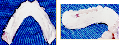
Denture space Impression after removal from the mouth
Its appearance is totally un-conventional. Any evidence of large areas of air entrapment, insufficient or excessive volume of impression material, or tray showing through necessitate re-taking the impression.
The Poured Denture space Impression-Four matrices are required to record the buccal, labial, lingual & ridge contours
The impression seated on the ridge matrix (with the buccal, labial and lingual matrices removed) is mounted against the upper cast on the articulator.
Silicone impression is then removed – buccal and labial matrices (surfaces) are replaced.
Softened wax is then placed in the space for setting the lower teeth for wax try- in.
The Waxed Trial Denture
The soft tissue contours are carefully developed without altering the basic contours of the recorded impression.
The routine assessments are conducted at the trial insertion, with special emphasis on the stability of the denture.
Some other techniques for recording Neutral Zone
Different designs of Impression trays
Injecting the Alginate into the Denture space ‘Impression tray is stabilized by biting’
Articulation & Set-up of teeth
Alginate impression acts as the index for tooth position
Replacing Impression material with Wax rim Setting the teeth with a plaster index
Further Applications of the Basic Technique
Determining the Fit of a completed denture to the Neutral Zone
Coat the polished surfaces of the denture with a low viscosity silicone impression material. Ask the patient to perform functional movements while the material sets. Inspect the denture & adjust any heavy muscle contact.
Determining the optimal space for a segment of the denture
Remove the teeth and the base material from the segment of the denture that needs modification. Apply adhesive and take the impression with moldable material. Check for stability and undertake the laboratory procedures.
Neutral Zone Versus Biometrics
Neutral Zone concept for the placement of artificial teeth could not enjoy the universal approval as did the Biometric concept of tooth arrangement. The reasons for this limited success are numerous, e.g.,
1. The viscosity of the material used for obtaining this impression is critical. More viscous the material, more it will be difficult for the muscles to mold it and visa versa.
2. The geriatric patients could suffer difficulty during the procedure as their musculature may have lost the tone.
3. The stability and retention of the bases on the soft denture support must be excellent as well as the comfort.
4. The resultant ‘neutral zone’ is often narrow and more lingually placed - with the closed mouth technique, the tongue could not perform all the functional movements, such as the phonetics.
5. This technique does not offer any guidelines for the selection of the teeth.
6. The technique is troublesome for the patient and does not offer much advantage over the biometric guides for the arrangement of teeth.
 Most of the patients seeking dental treatments might be having a problem of how to communicate with the dentist during the dental procedure. That is because our main source of communication is verble communication which will be affected during dental procedure.


