Wednesday, August 17, 2011

Treacher Collins syndrome-Discussion with a Clinical case history

  • This 4 years old female is a victim of Treacher-Collins syndrome.
  • Multiple facial anomaly including hypoplastic ears, anti-Mongolian eyes, mandibular facial dysosteosis , micrognathia, and choanal atresia were noted at birth.
  • Due to dyspnea and poor feeding, the patient was send to pediatric department.
  • Endo-trachea tube intubated for pneumonia was done at that time.
  • Because of extubation difficulty, tracheostomy was performed on 1987-11-28.
  • Distraction of mandible has performed twice on 1991-4-26 and 1991-11-29.
  • This time, for the purpose of remove tracheostomy, choanal plasty and stenting were arranged on1992-4-15.
History of the patient
Treacher-Collins syndrome with
     a. Micrognathia s/p distraction twice.
     b. bilateral microtia with hearing aid.
     c. PFO and PDA
     d. bilateral Choanal atresia s/p tracheostomy.
Family history: denied.
 
Treacher-Collins syndrome
·         What is treacher-Collins syndrome?
  • It is a condition in which the cheek bones and jawbone are underdeveloped.
  • Dr. Treacher Collins, a British ophthalmologist, was the first one who described the syndrome in 1900.
  • Franceschetti and Klein, who extensive reviewed the essential components of the condition, used the term mandibulofacial dysostosis to describe the clinical features.
  • Treacher Collins-Franceschetti syndrome 1 (TCOF1) was the other named of the syndrome.
Why and how does Treacher-Collins syndrome happen?
There are two possible ways that the syndrome develops:
                1. Develop as a new mutation.
                2. Inheriting it from one of the parents.
The incidence of the syndrome is about      1 : 10000~1 : 50000
Why and how does Treacher-Collins syndrome happen?
o   The syndrome is an autosomal  dominant disorder.
o   The gene mutated in this syndrome was mapped  at 5q32-33 initially.
o Two apparently balanced translocations, t(6;16)(p21.31;p13.11) and t(5;13)(q11;p11), and two interstitial deletions, del(4)(p15.32;p14) and del (3)(p23;p24.12)
Clinical features and diagnosis of the syndrome
  • The anomalies was due to the defects of the first and second branchial arches, clefts, and pouches during early embryonic development : 
  • Abnormalities of the pinnae which are frequently associated with atresia of the external auditory canals and anomalies of the middle ear ossicles. Bilateral Conductive hearing loss is common.
  • Hypoplasia of the facial bones, especially mandible and zygomatic complex.
  • Antimongoloid slanting of the palpebral fissures with colobomata of the lower eyelids and a paucity of lid lashes medial to the defect.
  • Cleft palate.


  • The clinic features are usually bilaterally symmetrical, and the non-penetrance is rare.
  • But due to the expression of the gene is extremely variable, the diagnosis and subsequent genetic counseling may be very difficult.
The minimal diagnostic criteria for the syndrome
  1. 40% of cases have a previous family history.
  2. Cranio-facial radiographs, particularly the occipito-mental view.
  3. Chromosome and gene exam.
What problems can be expected?
The most common difficulties involve:
  • Breathing
  • Hearing
  • Vision
The reasons that may cause breathing problems:
  • Small or undeveloped jaw
  • Cleft palate and choanal atresia
  • Tongue drop
  • Pharyngeal hypoplasia
  • Dyspnoea while developing colds and infections due to congestion and swelling of the airway.
  • Sleep apnea – affect the child’s mental development.
  • Speech therapy was needed.
The role of an anesthesiologist
This doctor is a very important part of any craniofacial team. Children with craniofacial problems often have problems associated with the airways that create breathing difficulties. It is essential that this doctor be well trained in pediatric anesthesiology, but it is just as important that he/she have substantial experience in dealing with these special children. The pediatric anesthesiologist’s amount of experience with craniofacial problems perhaps has the greatest effect on the overall safety of the surgery.

Thursday, August 11, 2011

Snake Bites Types,Diagnosis and Management-Medical Lecture Note


            3500   Species
            300     Venomous
            30000 - 40000 Death annually
  o   India
  o   Brazil
  o   Buruma

Poisonous Snakes
5 families
  1. Crotalidae-rattle snake,pit viper
  2. Viperidae-russel’s ,sawscaled viper
  3. Elapidae-cobra,krait
  4. Hydrophiodae-sea snake
  5. Colabridae 

    Indian scenario
    5 dangerously poisonous snakes
    ·         king cobra
    ·         common cobra
    ·         common krait
    ·         russell’s viper
    ·         sawscaled viper

    Most common poisonous snake is common krait
    ·         216  species
    ·         52  venomous
    ·         Ireland
    ·         Newzealand

    Snake venom
    ·         Toxic saliva secreted by modified parotid glands of a venomous snake
    ·         Amber coloured when fresh
    Constituents              
    ·         Toxins
    ·         Enzymes
    ·         Neurotoxins
    ·         Cardiotoxins
    ·         Hemolysin

    Venom apparatus

    Fangs

    Venom classification
    ·         Neurotoxic-Elapids(cobra,krait)
    ·         Hemotoxic-Viperidae
    ·         Myotoxic-Sea snake

    Krait and russell’s viper is much more toxic than that of cobra
    Symptomatology of non venomous snakes
    ·         Universal fear associated induce a state of shock
    ·         Bite site may demonstrate multiple teeth impressions
    ·         Lack of significant local pain or swelling
    ·         Adequate reassurance and symptomatic treatment measures lead to full recovery

    Symptomatology of venomous snakes
    1.       Elapid bite
    Local features:
    ·         Indistinct fang marks ,
    ·         Burning pain,
    ·         Swelling and discolouration,                                                                         
    ·         Serosanguinous discharge
           
    Systemic features
    ·         Preparalytic stage: Emesis,headache, LOC.
    ·         Paralytic stage: ptosis,ophthalmoplegia drowsiness,dysarthria, dysphagia,convulsions, bulbar paralysis, resp failure .

    2.       Viperid bite
    Local features:
    ·         Rapid swelling,
    ·         Discolouration,
    ·         Blister formation,
    ·         Bleeding from bite site,
    ·         Severe pain
        
    Generalised bleeding manifestations.                       
    • epistaxis,
    • hemoptysis,
    • bleeding gums
    • hemauria
    • purpuric spots
    • Renal failure

    Hydrophid bite
    Local features:
    §  minimal swelling and pain
    Systemic features:
    §  Myalgia muscle stiffness
    §  Myoglobinuria ,
    §  Renal tubular
    §  Necrosis                  

    Diagnosis of snake bite
    1.       Fang marks:classically, two puncture wounds seperated by a distance varying from 8mm to 4cm, depending on the species involved.
    2.       However a side swipe may produce only a single puncture,while multiple bites could result in numerous fang marks.
    3.       Bailey’s method
      
    Identification  of snake
    §  Poisonous  or non poisonous
    §  Species
           Venomous                  Non venomous
    Stout, dull coloured             abruptly tapering tail
    Slender,brightly coloured gradually tapering tail
    Tail: may be rounded or flattened
    Always rounded
    Belly scales are broad
    Belly scales are small..do not extend the entire width
    Head :triangular
    Rounded or oval
    Head scales: usually small
    Usually large (shields)
    At least one pair of teeth in the upper jaw are modified to form fangs
    All teeth are uniformly small in size
    Saliva contains toxic peptides and enzymes
    Non toxic


    Non-venomous snakes

    Kurudan
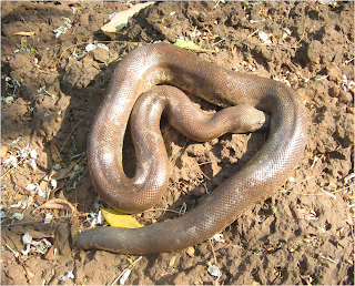
    Red Sand Boa
    Eryx conicus (chenathandan)
    Python
    Bronze back tree snake
    Ckeckered keelback


    Striped keelback
    Cat Snak


    The Venomous snakes


    Banded  krait
    Hook nosed sea snake

    Pit viper


    Saw Scaled viper

    Slender coral snake
    Management of snake bite
    1.       First aid treatment
    2.       Transport to hospital
    3.       Rapid clinical assessment and resuscitation
    4.       Investigations/laboratory tests
    5.       Antivenom treatment
    6.       Supportive/ancillary treatment
    7.       Treatment of the bitten part
    8.       Rehabilitation
    9.       Treatment of chronic complications

    First aid
    §  Delay enry of venom
    §  Tourniquet
    §  Above knee
    §  Above elbow

    Useless or Dangerous Methods
    §  Making local incisions or pricks at the site of the bite or in the bitten limb
    §  Attempts to suck the venom out of the wound
    §  Use of snake stones
    §  Electric shock
    §  Topical instillation or application of chemicals, herbs or ice packs.

    Clinical assesment
    Vital signs
    o   pulse
    o   BP
    o   Respiration –SBC

    Observe
    o   Bite mark
    o   Local reaction
    o   Painful INE
               
    Neurotoxicity
    o   Ptosis
    o   Ophthalmoplegia
    o   Myasthenia like symptoms
    o   Asses SBC
    Haemotoxicity
    o   Purpura
    o   Echymosis
    o   Gingival sulcus bleed
    o   Hematuria
    Capillary leak syndrome
    o   Puffiness
    o   Chemosis
    o   Parotid swelling

    Lab. Investigations
    Haematological
    o   Leucocytosis(>20,000-severe envenomation)                          
    o   Elevated PCV
    o   Thrombocytopenia
    o   Evidence of hemolysis
    o   Prolonged ct,pt,ptt
    o   Elevated fdp
    Ct     >    20 minutes
    Sure sign of envonomation
    Pitviper   > 2 weeks
    ECG: bradycardia
                 ST-Elevation or other way
                 T-wave inversion
                 QT Prolongation
                 Changes due to hyperkalemia
                 
    Metabolic
             Hyperkalemia
             Hypoxemia with resp.ac
             Met.ac or lactic ac
    Urine
             Hematuria,proteinuria,Hburia
             Mburia
            
    Renal : ARF -- BU S.Cr   S E
    CXR :  Pulm.edema
               Intrapulm.hgs
               Pleural effusion
    Immuno-diagnosis: by ELISA….
    Highly sensitive but specificity inadequate to diff b/w diff species of snakes
    Specific management  asv
    Horse serum
    Asv in india
    o   cobra
    o   krait
    o   russel’s viper
    o   saw scaled viper

    1 mi ASV     -   0.6mg cobra R viper
                        -   0.45mg krait S viper
                           
    Indication -Systemic Manifestaiton
    o   Neurotoxicity
    o   Repeated vomiting
    o   Haemotoxicity
    o   Nephrotoxicity
    o   Cardiotoxicity
    o   Rhabdomyolysis

    Prolonged CT  alone
    o   Pit viper  -  no
    o   Snake not identified

    Neurotoxic envonomation
    o   Initial dose  10  -  15 vials
    o   Reassess
    o   Improvement   30  --  60 min
    o   Repeat  5 vials after 60  -- 90 mins
    o   Supportive – neostigmine after atropine

    Haemotoxic envonomation
    Mild            CT < 30 mins
                       Clot size = 50% blood col
                       Initial dose = 5 vials
    Moderate   CT   > 30 mins
                       Clots only speckles
                       initial dose   = 10 vials
    Severe      incoagulable
                     Initial dose  = 15 vials
    Repeat ct after 6 – 9 hours
    If ct pronged repeat 5- 10 vials
    Low dose infusion – following days
    Supportive care
    o   Antibiotics
    o   Methyl prednisolone
    o   FFP, fresh blood
    o   Prevention and rx of hypotension
    o   prevention of shock
    Prevention of arf
    ·         Proper fluid administration
    ·         Correct myocardial dysfunction
    ·         Monitor output bu s.cr se                
    ·         Avoid  nephrotoxic drugs
    ·         Protein restriction

    Management of local reactions
    ·         Bullae   - left intact
    ·         Necrosis  - debridement
    ·         Compartment syndrome – fasciotomy
    ·         Most comfortable position

    Reactions to anti venom
    ·         Anaphylactoid 10 – 90 mins
    ·         Pyrogenic 2 hours
    ·         Serum sickness 5 – 21 days

    Bee and wasp sting
    ·         Neurotoxic
    ·         Hemolytic
    ·         Hypersenstising agents - anaph
    ·         Rx  local antihistamines

    Scorpion sting
    ·         Hemo / neuro toxic
    ·         Sting mark one hole in centre
    ·         Take ecg to r o cardiotoxicity
    ·         Rx local anaesthetic ice pack

    Popular Posts

    Join This site