Thursday, October 27, 2011

Maxillary Sinus in Health and Disease-Oral Surgery Lecture

Anatomical facts and location:
√ The largest para-nasal sinuses.
√ Situated in the maxilla.
√ Has pyramidal shape.
√ Lateral nasal bone forms its base.
√ Apex headed towards the zygomatic bone.
√ Canine fossa, orbital floor and hard palate form the pyramidal walls.
√ Communicates with nasal cavity through maxillary ostium, in the posterior end of hitus simlunaris of middle meatus.


Anatomical morphology:
√  Size varies from one person to another.
√  Asymmetry existed in the same individual.
√  Small in children and grows up with aging.
√  Average height is about 3.5 cm, depth 3.2 cm and width 2.5 cm.
√  Capacity of about 15 cc.
√  Divided into several compartments by bony septa (underwood’s septa).
√  Lined with pseduo-stratified columnar ciliary epithelium (schneiderian membrane).


Relation with other structures:
√ Alveolar bone and dentition.
√ Nasal cavity and nasopharynex.
√ Orbital cavity and its contents.
√ Hard palate and oral cavity proper.
√ Pterygomaxillary fissure and its contents.
√ Neurovascular structures including infraorbital and superior alveolar nerve.


Development:
√  Develops from invagination of the mucous membrane of middle meatus of the nasal cavity at about the 3rd month of intrauterine life.
√  Fully development reaches with the age of 16 years.
√  Loss of permanent teeth and alveolar bone may make the sinus to appear huge in size.

Blood supply:
Blood supply from facial, maxillary, infraorbital, greater and lesser palatine arteries and lateral and posterior nasal branches of sphenopalatine artery.
Venous drainage to the anterior facial vein, sphenopalatine vein and pterygopaltine plexus.


Nerve supply:
√  Infraorbital nerve.
√  Posterior, middle and anterior superior alveolar nerves.
√  Greater and lesser palatine nerves.


Lymphatic drain:
The lymphatic drain of the sinus is through the nose or the submandibular lymph nodes.

Physiology:
Unknown but the following functions have been proposed:
√  Speech and voice resonance.
√  Reduce weight of skull.
√  Warmth inspired air.
√  Filtration of inspired air.
√  Immunologic barrier ( body defense).

Pathology:
  1. Congenital anomalies.
  2. Inflammatory diseases.
  3. Cysts and odontogenic infection.
  4. Bone metaplasia and benign tumors.
  5. Neoplasia.
  6. Trauma.


Congenital anomalies:
√  Cleft palate.
√  Facial fistula and cleft.
 √ Cystic formation.
√  Atresia.

Inflammatory diseases:
√  Bacterial infection.
√  Bacterial infection secondary to viral infection.
√  Fungal infection.

Sinusitis

Acute sinusitis:
Suppurative or non suppurative inflammation of the mucosal lining of the sinus. It involves one or both sinuses.

Causes:
√  Secondary to hay fever and allergic rhinitis.
√  Secondary to acute rhinitis (common cold) and URT infection.
√  Bacterial infection due to: dental sepsis, swimming and diving, trauma and foreign body dislodgment.

Sings and symptoms:
√  Headache.
√  Pain and tenderness.
√  Nasal obstruction.
√  Nasal discharge.
√  Toxic manifestations.
√  Heavy filling with bending.
√  Nasal congestion.
√  X-ray and transillumination findings.

Treatment:
√  Rest and fluid and mouth hygiene.
√  Antibiotics (C&S); pneumococci and streptococci are the most causative organisms.
√  Analgesics and antihistamines.
√  Local treatment (decongestant and steam inhalation).
 
Chronic sinusitis:
It is a chronic type of infection affected the mucosal lining of one  or both sinuses, resulted in mucopus or pus collection. A polypoidal type of inflammation can lead to formation of multiple or single mucosal polyps. 

Causes:
√  As a consequence of non resolved acute sinusitis.
√  Dental abscesses.
√  Virulent organism with low resistance.
√  Foreign body dislodgement or trauma

Signs and symptoms:
√  Headache.
√  Nasal obstruction
√  Nasal discharge.
√  Fatigue.
√  Hyposmia/ cacosmia.
√  Transllumination findings.
√  Proof puncture.

Treatment:
√  Antibiotics.
√  Systemic decongestants.
√  Sinus wash-out.


Mycotic infection:
Aspergillosis:
Opportunistic infection caused by maxillary sinus flora fungi environment in susceptible individual, leads to obliteration of the sinus space and erosion of its bony components.

Complications of sinusitis:
  • Orbital abscess and orbital cellulites.
  • Intracranial abscesses.
  • Meningitis.
  • Cavernous sinus thrombosis.
  • Spread of infection to neighboring sinuses, structures and organs.
  • Osteomyelitis.
  • Gastrointestinal disturbances.

Cysts and odontogenic tumors:

Odontogenic cysts:
√ radicular cysts.
√ residual cysts.
√ dentigerous cysts.
√ premordial cysts.

Non-odontogenic cysts.
Mucocele and retention cysts.

Odontogenic tumors:
√ ameloblastoma.
√  Myxoma.

Bone metaplasia and benign tumors:
√  Fibrous dysplasia.
√  Ossifying fibroma.
√  Transitional papilloma.
√  Osteoma.
√  Giant cell lesions.

Neoplasia:
√  Squamous cell carcinoma.
√  Adenocarcinoma.
√  Sarcoma (osteosarcoma).
√  Ewing’s sarcoma.


Trauma:
√  Tuberosity fracture.
√  Dentoalveolar fracture.
√  LeFort’s fractures.
√  Zygomatic complex fracture.
√  Pure and impure orbital floor fractures.
√  Establishment of oro-antral fistula.

Clinical examination:

Inspection
√ Assess asymmetry.
√ Color of overlaying skin.

Palpation
√  Tenderness.
√  Swelling and expansion.
√  Depression.


Examination of nasal passage
√ Nasal patency.
√ Pus discharge.
√ Nasal polyps.
√ Erythema, redness, change in the color of nasal mucosa.


Transillumination 
Diagnostic sinus lavage
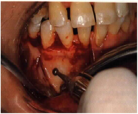
√ sinus rinsing through the canine fosaa.
√ Nasal antrostomy.


Radiographical examination:
Routine radiographical examination
√ Orthopantomogram (OPG)
√ Occipitomental (water’s view), with lateral tilt.


Special investigation and radiographical examination
  • Sinuscopy
  • Sinogram
  • CT scan
  • MRI
  • Microbiology and histological examination:
  • Culture and sensitivity and biopsy.


Wednesday, October 26, 2011

Diagnosis and Management of Hemorrhage in Oral Surgery

What is meant by Hemorrhage ?
Prolonged or uncontrolled bleeding is often referred to as hemorrhage.
The amount of blood lost as a result of hemorrhage can range from minimal to significant quantities.

Hemorrhage in Surgery
Hemorrhage  can occur to a greater or lesser degree during all surgical procedures and it’s management depends upon whether the patient is hematologically normal or suffers from some disturbance in the normal clotting mechanism.

The overwhelming majority of patients who undergo oral surgical procedures are those who have normal haemostatic mechanism.
Therefore, significant or major hemorrhages are not that common in oral surgery except in patients who have a bleeding / clotting disorder or those who are on anticoagulants.

However, uncontrolled and persistent bleeding can occur in some healthy patients after dental extraction.
Therefore, it is still important to achieve proper hemostasis in all patients during oral surgical procedures, so as to prevent excessive post-operative blood loss.

Normal Mechanism of Hemostasis
Hemostasis is a complicated process.
It involves a number of events

Hemostasis - Normal Mechanism
1.  Vascular phase
2.  Platelet phase
3.  Coagulation phase

Vascular phase
When a blood vessel is damaged, vasoconstriction results.

Platelet phase
Platelets adhere to the damaged surface an form a temporary plug.
Through two separate pathways, the Intrinsic and Extrinsic, the conversion of fibrinogen to fibrin is complete. Fibrin tightly binds the platelets to form a clot

Coagulation phase

The clotting mechanism


Hemostasis
Dependent upon:
  • Vessel Wall Integrity 
  • Adequate Numbers of Platelets 
  • Proper Functioning Platelets 
  • Adequate Levels of Clotting Factors 
  • Proper Function of Fibrinolytic Pathway
Hemorrhage in Oral Surgery
Hemorrhage following Oral Surgical procedures can occur due to local or systemic causes.
In healthy patients the postoperative bleeding is mainly due to local causes.

Local causes of hemorrhage in oral surgery
Local causes of hemorrhage originate in either soft tissue or bone.

Local causes of hemorrhage in oral surgery –Soft tissue bleeding
Soft tissue bleeding is either arterial, venous, or capillary in nature.

Arterial bleeding is bright red and spurting in nature.
Arteries in the soft tissues at risk during oral surgical procedures are the lies posterior portion of hard palate) greater palatine artery and the buccal artery (lies lateral to the retromolar pad)

Venous blood is dark red in color and flows steadily and heavily especially if the vein is large.

Capillary bleeding is bright red in color and is more of a minimal ooze.

Local causes – Osseous (Bony) bleeding in oral surgery
Troublesome bone bleeding originates either from nutrient canals in the alveolar region, central vessels, such as the inferior alveolar artery, or from central vascular lesions (Hemangioma or Vascular malformation)

Systemic causes of hemorrhage in oral surgery
Some patients with heriditary conditions such as hemophilia, Von Willebrand’s disease are susceptible for hemorrhage following oral surgical procedures.
Patients with thrombocytopenia (decreased platelet count) , Leukemia e.t.c., are also at risk of prolonged bleeding after surgery.

Patients with uncontrolled hypertension.
Patients with H/O prosthetic heart valve replacement, Stroke (Cerebrovascular accident) e.t.c., take oral anticoagulants like Aspirin or Warfarin to prevent the occurrence of a thromboembolic episode.
These patients are also at risk of prolonged severe bleeding during and after an oral surgical procedure.

Types of Hemorrhage - Primary Hemorrhage
This occurs during the surgery, as a result of injury like cutting or laceration of the  artery or bleeding from bone.
This also occurs when surgery is done in an infected area with a lot of granulation tissue.
It can also occur after a very short period of time  immediately after surgery.
This type of bleeding is really normal and can be controlled easily.

Types of Hemorrhage - Intermediate / Reactionary Hemorrhage
This type of bleeding occurs within a few hours after surgery.
This type of bleeding occurs as a result of failure of coagulation to occur (as in patients with systemic bleeding problems or those on anticoagulants)
Patients who have unknowingly disturbed / dislodged the clot are also prone for this type of bleeding.

Types of Hemorrhage - Secondary Hemorrhage
This occurs after 7 to 10 days after surgery. This is mainly due to partial division of blood vessel in combination with infection of the wound (Like patient’s who undergo radical neck dissection e.t.c.,).
This type of bleeding is not very frequently encountered after oral surgery procedures.

Management of Primary Hemorrhage in Normal patients
The management of bleeding during surgery (Primary bleeding) can be achieved by the following means,
  • Securing / ligation of blood vessels with silk sutures. 
  • Use of pressure swab to achieve hemostasis. 
  • Use of electrocautery to achieve hemostasis. 
  • Use of hemostatic agents like bone wax, surgicel,e.t.c., 
  • Hypotensive anaesthesia (G.A) and use of vasoconstrictors in L.A.
Local Measures  ( Synthetic Materials)
There are several materials that are commercially available that are used  locally for achieving adequate hemostasis.

Local Measures: Surgicel (Oxidised Regenerated Cellulose)


Local measures: Gelfoam with activated thrombin


Local Measures: Avitene (Microfibrillar Collagen)


Local Measures: Etik Collagen (Packed collagen)


Local Measures: Tranexamic acid 5%


Local Measures: Tranexamic acid 5% in Syringe


Local Measures: Irrigation of wound with Tranexamic acid


Local Measures: Suturing the wound


Local Measures: Pressure with oral packs


Management of Intermediate Hemorrhage in  Normal patients

The management of bleeding that occurs immediately after surgery (Reactionary bleeding) involves proper examination of the surgical wound to identify the site of bleeding (i.e ) from bone or soft tissue.
If bleeding is from bone then the hemostatic agents like bone wax or gelfoam is usually used.
If bleeding is from soft tissues then, ligation / cauterization of blood vessels along with the use of hemostatic agents like surgicel and suturing of the wound is carried out.

Management of Secondary Hemorrhage in  Normal patients

The management of this type of bleeding that occurs a few days after surgery involves the removal of any debris from the wound surface that promotes the infection of the wound.
Identify the source of bleeding and treat as would be done in a  patient with secondary bleeding.
Surgical stents can be placed over extraction sockets for stabilization of clot and prevention of  wound contamination.

Management of Hemorrhage in patients with  bleeding disorders / and those on anticoagulant therapy

The usual protocol involved in the treatment of this group of patients consists of pre-operative blood investigations and preoperative correction of the underlying deficiency (Replacement of Clotting factors / platelets) if any in these patients.
Subsequently, after this appropriate local measures are used to decrease the chances of post-operative bleeding.
 
Laboratory evaluation
  • Platelet count 
  • Bleeding time (bt) 
  • Prothrombin time (pt) 
  • Partial thromboplastin time (ptt) 
  • Thrombin time (tt)
Platelet count
Normal  100,000 - 400,000 cells/mm3 
< 100,000            Thrombocytopenia
50,000 - 100,000 Mild Thrombocytopenia
< 50,000              Severe Thrombocytopenia
                       
Bleeding time
Provides assessment of platelet count and function
Normal value
2-8 MINUTES

Prothrombin time
Measures Effectiveness of the Extrinsic Pathway
Normal value
10-15 SECS

Partial thromboplastin time
Measures Effectiveness of the Intrinsic Pathway
Normal value
25-40 SECS

Thrombin time
Time for Thrombin To Convert  Fibrinogen to Fibrin
A Measure of Fibrinolytic Pathway
Normal value
9-13 SECS

Management of Hemorrhage in patients with uncontrolled hypertension.
This group of patients need appropriate medical consultation for initiation of medical treatment to decrease their Blood Pressure.
Thus once their B.P is controlled, then the bleeding decreases and with local measures the hemorrhage is controlled.


Surgical management of Unerupted and Impacted teeth

Terminology
  • Unerupted tooth (retained tooth): is that fail to erupt into the oral cavity at the normal time and age.
  • Impacted tooth: is a retained tooth that is completely or partially buried in the soft tissue or the bone.
  • Aberration: is a tooth that develop distant from its normal location. 
  • Ectopic eruption: eruption of a tooth outside the arch line based on clinical evaluation.
  • Agenesis: failure of a tooth to develop due to many reasons and genetic factor is highly contributed.
 Common unerupted and impacted teeth:
  1. Mandibular third molars
  2. Maxillary canines
  3. Mandibular second premolars
  4. Maxillary second premolar
  5.  Mandibular canines
Etiology of failure of eruption
  1. Tooth agenesis.
  2. Injury to tooth germ and displacement of tooth follicle.
  3. Crowding and disproportion between teeth size and jaw.
  4. Premature loss of a deciduous predecessor and gingival fibromatosis
  5. Presence of supernumerary teeth
  6. Presence of tumors or cysts
  7. Cleft palate and alveolus
  8. Cleidocranial dysostosis
  9. Conginital brevicollis dystrophy
  10. Klipped feil syndrome
  11. Hypopituitarism
  12. Cretinism (infantile hypothyrodism)
  13. Rickets
Indications-(Rational for treatment)
  • Majority removed because of pain or being a foci of infection.
  • Involvement in pathology like cyst and tumors.
  • Resorption of roots of adjacent teeth.
  • Interference in line of osteotomies and fractures.
  • Infection of surrounding soft or hard tissue.
  • For prophylactic reasons.
Contraindications-(Relative)
Asymptomatic unerupted teeth that removal is possibly complicated by an injury to inferior dental or lingual nerve during surgery treatment.
Teeth of favorable position that can be monitored at time intervals to detect the development of any complications.

Recognition of the problem:
The existence, position, orientation of the impaction and diagnosis of associated problems are based on:
  • History
  • Clinical examination
  • Radiography
History
Missing tooth or teeth with or without history of pain and swelling of underlying mucosa (agenesis??)
In case of pain, effort must be paid to eliminate other possible causes of dental pain from another tooth such as pulpitis and periodontitis.
Pain at posterior aspect of the mouth that can be a refereed type of pain such as earache, eye pain, artherolgia, etc.)
Inflammation around the crown of the tooth that make more acute symptoms (pericoronitis).

Examination
  • Recording of missing teeth.
  • Recording of retained deciduous teeth.
  • Identify caries and periodontal diseases.(pain might be from adjacent carious tooth, this would influence the proposed treatment planning)
  • Vitality test of all teeth in doubt.
  • Examination for sign of infection.(swelling, discharge, trismus and enlarged lymph nodes)
  • Facial asymmetry and jaw bone expansion.
Radiography-(objective indications)
  • To disclose the unerupted tooth and the texture of the surrounding bone.
  • To disclose the position in the jaw and its relation to adjacent teeth and other vital structures (sinuses, IDC,).
  • To disclose the crown-root ratio and roots configuration.
  • (curvature, numbers, hypercemntosis, bulbous, fused or diverged)
  • To disclose the degree and orientation of impaction.
  • To disclose atrophy of dental follicles and existence of pathological development.
Preoperative assessment
Asymptomatic unerupted teeth most often discovered following radiographical screening (accidental findings).
Partially erupted teeth might be associated with pain and infection.
Impacted lower wisdom tooth may cause crowding upon anterior teeth.
Impacted tooth may erode or cause cavitation of adjacent teeth.
Impacted teeth may be associated with pathological cyst development.

Pericoronitis
  • Infection involves the soft tissue surrounding the crown of partially erupted tooth.
  • Usually caused by streptococci and anaerobic bacteria.
  • It may presented as an acute or chronic infection.
  • Acute infection developed over hours and days and associated possibly with systemic manifestation.
  • Chronic infection distinguished by redness and or discharge of pus with few acute symptoms lasting over weeks to months.
  • It may be associated with poor oral hygiene and upper respiratory infection.
Signs and symptoms:
  • Swelling of retro-molar tissue
  • Soreness
  • Erythemia of overlaying soft tissue or operculum
  • Trismus
  • Facial swelling of the affected side
  • Raised temperature
  • Regional lymphodenopathy
  • General malaise
Contributory factors:
  • Trauma from an opposing over-erupted wisdom tooth
  • Entrapment of food debris and bacterial infection under the operculum
  • Physical and mental stress
  • Pregnancy and suppression of the immune system
  • Upper respiratory tract infection
Management of pericoronitis
  1. Local irrigation by hot salt mouthwash. chlorhexidine mouthwash
  2. Antibiotics if signs of spreading infection are evident. (amoxycillin, metronidazole)
  3. Analgesic and non-steroidal anti-inflammatory agent.
  4. Extraction of upper opposing wisdom tooth if traumatizing the lower operculum.
  5. Removal of lower wisdom tooth when acute infection is resolved.
  6. Hospital admission in case of severe infection that may compromise the airway.
Considerations in clinical examination of an impacted/ partially erupted tooth
  1. Patient age and tooth eruption
  2. Associated infection
  3. Caries and restoration
  4. Dental status of the adjacent tooth
  5. Periodontal status
  6. State of the TMJ
 Status of tooth in question
  • Based on clinical evaluation
  • Erupted but non-functional (no opposing, tilted, carious, etc.)
  • Partially erupted (covered partially with soft tissue)
  • Partially erupted with sign of recurrent infection
  • Truly impacted (bony or soft tissue)
  • Association with pathological lesions
Methods of radiographical examination
Radiographs in two planes at right angles are needed to show clearly the position of the tooth and the degree of impaction
  • Orthopantomogram (OPG)
  • Preapical radiograph
  • Lateral oblique view of the jaw
  • Vertex occlusal view
  • Parellex method of Clark
Radiographical assessment
  • Orientation (mesioangular, vertical, distoangular and transalveolar)
  • Depth below the occlusal plane
  • Crown size and follicular width
  • Root morphology (number, length, shape: fused or separate, curved apex, bulbous, ankylosis).
  • Condition of the crown and the adjacent tooth
  • Approximation of an ascending ramus, IDC, maxillary sinus, ptrygoid plates and pyriform fossa.


Management of impacted/ partially erupted teeth
 

Options of management 

  1. No treatment
  2. Conservative management
  3. Surgical repositioning and transplantation
  4. Exposure of the teeth with or without orthodontic application
  5. Surgical removal

No treatment-(Choices of putting tooth in probation)
  • Asymptomatic tooth
  • When it acts as a buttress for the root of adjacent tooth
  • When vital structures are at risk of injury in the course of operation
  • In case of acute preicoronitis

Conservative management
  • Tooth that might be brought into occlusion provided that space is adequate in the arch line.
  • When adjacent tooth is carious, heavily filled or missing.
  • Mesial drifting may allow tooth to replace poorly prognosis or missing anterior one

Surgical repositioning and transplantation
  • Aimed to move tooth bodily into the dental arch
  • Careful surgical extraction is required to minimize the damage to the apical vessels and periodentium
  • Imobilization within the prepared socket for 4 weeks
  • Success determined by the dental age (unclosed apices), patient age and atrumatic surgery
  • Resorption of root might be evident in 2-5 years
  • Early endondontic treatment might be of help to minimize the failure


Third molar transplantation 


Surgical aids to orthodontics
  • It is mostly prescribed for impacted canine
  • Other teeth might be considered as well
  • Aimed to help in establishing optimum occlusion orthodontically
  • The canine is very important esthetically
  • The success is very high
  • Surgery for exposure is much easier than for removal of the impaction
Surgery-assisted orthodontic traction
  • Reflection of mucoperiosteal flap
  • Crown is to be freed to its greatest circumference
  • Preservation of attached ginigiva for labially and buccally placed teeth
  • Orthodontic device ( button, hock and ligature wire) is to be applied
  • Flap is to be then sutured  back in position
  • For palatal placed teeth, soft tissue excision for exposure is to be packed with whitehead’s varnish, BIPS, coepack
  • Orthodontist visit to be arranged one week post op for traction application
  • The procedure:


Removal of unerupted teeth
  • Earlier to sclerosis of bone
  • Earlier to follicle atrophy
  • When it is infection-free
  • Before fully development of roots
  • When 2/3 of the roots are formed
  • Best timing for removal
 Surgical Considerations
  • Localization of unerupted tooth
  • Morphology of the tooth and roots
  • Relationship to the inferior dental neurovascular bundle
  • Buccolingual position
  • Relationship to adjacent teeth
  • Relationship to inferior border of the mandible and anterior border of the ramus
Planning for operation
  • “Reverse in order”
  • The tooth position in jaw
  • The natural line of withdrawal
  • Overcome obstacles (ascending ramus and adjacent tooth or teeth)
  • Point of application for elevation
  • Access by removing bone and design flap accordingly
Natural line of withdrawal
Teeth extracted by moving them away from sockets or bone along their pathway
The course of movement is dictated by the curvature of the roots
Unfavorable elevation refers to tooth goes deeper in bone or impacted against another tooth

Violation of the principles of line of withdrawal
Fracture of bone (the whole entity or part of it)
Displacement of tooth into soft tissue or anatomical spaces
Damage of inferior dental nerve
 
Obstacles to elevation
Intrinsic
√ shape of the tooth and root
√ Constriction at the neck of the tooth
Extrinsic
√ bone and depth of the tooth
√ adjacent tooth (impaction against a tooth )
√ adjacent vital structures (the inferior dental neurovascular bundle)

Overcoming the obstacles
Removing sufficient bone to allow tooth to be rotated and delivered
Division of the tooth horizontally or vertically or by both using:
√ drill and large fissure bur
Removal of lingual plate using:
√ chisels and mallet
 
Point of application
Dental elevators is the best for removal of buried teeth
Point of application must be determined during planning
Point of application is to be prepared simultaneously during access preparation
No tooth division until adequate point of application has been prepared.

Preparation for surgery
Hospital and general anesthesia
Outpatient clinic with either intravenous sedation or local anesthesia

Surgical access
  • Flap must be sufficient enough to allow direct vision with no chance of tension and trauma
  • Bone removal should permit tooth with its greatest crown dimension to pass freely (tooth division may minimize the need for more bone removal)
  • Curved and bulbous root must be made free of bone
  • Cutting of bone and tooth division must be completed before attempting elevation
  • Flap is to be replaced and rests on bone before suturing
Closure of wound
  • Debridment and smoothening of sharp edges of the socket
  • Removal of dental follicle (sack) without endangering vital structure (lingual nerve)
  • Primary closure as long as flap is not under tension is desirable
  • Resorbable or non-resorbable suture may be used
  • Suture notes should be kept to a minimum
Surgery of Mandibular Impacted/ Parially Erupted Teeth


Position
  • Vertical
  • Horizontal
  • Mesioangular
  • Distoangular
  • inverted
  • Transbuccally (crown facing lingually or buccally)
  • Apparent position; ramus or close to inferior border of the mandible


Impacted mandibular canine and premolar tooth
 

Maxillary Teeth
  • Surgical considerations
  • Position of unerupted tooth (3rd molar or canine)
  • Relationship to adjacent teeth
  • Relationship to maxillary sinus
  • Morphology of the roots
  • Status of adjacent teeth
  • Presence of supernumerary and supplemental teeth

Upper third molar Operative technique
  • The flap
  • The envelop flap
  • Two sided flap (triangular type)
  • Bone removal
  • Establishment of OAF
  • Closure

Surgical exposure and surgical removal of impacted canine and anterior

Palatally positioned tooth

Labially positioned tooth


Removal of unerupted teeth from edentulous ridge
surgical consideration
  • Difficulty is owing to sclerotic bone and loss of periodontal space
  • Gentle force via a well prepared point of application would minimize the risk of fracture of brittle bone
  • Alveolar ridge preservation by accurate assessment and minimal bone removal
  • Osteoplastic flap to preserve the alveolar bone in height and in width
  • Bone reduction and fixation in the incident of atrophic jaw fracture

Difficulties-associated surgery
  • Small mouth
  • Narrow space between anterior border of the ramus and distal aspect of second molar tooth buried deeply in bone
  • Approximation of inferior dental canal and sinuses
  • Existence of fusion and ankylosis
  • Devitalizations and cavitations

Complications associated with unerupted and impacted teeth surgery
Intraoperative:
√ hemorrahge
√Fractured root, tuberosity
√Damage to adjacent tooth, tooth displacement
√ oroantral-oronasal communication
√Fracture mandible
Postoperative:
√ pain, swelling, bruising, trismus, aneathesis, infection


Popular Posts

Join This site