Saturday, October 19, 2013

Pulp therapy for primary teeth


Primary objective of pulp therapy is to maintain the integrity and health of the teeth and their supporting tissues by maintaining the vitality of the pulp of a tooth affected by caries, traumatic injury or any other cause damaging the liveliness of the pulp.
Indication and the type of the pulp therapy depends on the status of the pulp, whether it’s nonvital or vital and the type of the tooth whether its primary, young permanent or permanent. Status of the pulp would be determined by clinically with a proper history and a thorough clinical examination and by accurate special investigations such as vitality testing and radiographs. In this article I would mainly consider on the treatments to the pulp in primary teeth.


Vital pulp therapy for primary teeth diagnosed with a normal pulp or reversible pulpitis

Indirect pulp treatment
A procedure performed in a tooth with a deep carious lesion approximating the pulp but without signs and symptoms of pulp degeneration. The caries surrounding the pulp is left in place to avoid pulp exposure and is covered with a biocompatible material. A radiopaque liner such as a dentin bonding agent, resin modified glass ionomer, calcium hydroxide, zinc oxide-eugenol or glass ionomer cement is placed over the remaining carious dentin to stimulate healing and repair. Then the tooth is restored with a material that seals the tooth from micro leakage.


Direct pulp treatment
When a pinpoint mechanical exposure of the pulp is encountered during cavity preparation or following a traumatic injury a biocompatible radiopaque base such as mineral trioxide aggregate (MTA) or calcium hydroxide may be placed in contact with the exposed pulp tissue. Finally the tooth should always be restored with a material that seals the tooth from micro leakage.


Pulpotomy
A pulpotomy is performed in a primary tooth with extensive caries but without evidence of radicular pathology when caries removal results in a carious or mechanical pulp exposure. The coronal pulpotomy is amputated and the remaining vital radicular pulp tissue surface is treated with a medicament such as Buckley’s solution of formocresol. Gluteraldehyde and calcium hydroxide have been used but with less long term success. MTA is a more recent material with a high rate of success in pulpotomies. The coronal pulp chamber can be filled with zinc-oxide eugenol or other suitable base followed by acoronal restoration to avoid micro leakage and failure of the treatment. The most effective long term restoration has been shown to be a stainless steel crown although other alternatives such as composite resin and amalgam play a role when an adequate amount of enamel is intact.


Nonvital pulp therapy for primary teeth diagnosed with irreversible pulpitis or necrotic pulp

Pulpectomy
This involves the complete amputation of the pulpal tissue in a tooth that is reversibly infected or necrotic due to caries or trauma. The root canals are debrided mechanically with hand or rotary files and chemically with disinfectants such as sodium hypochlorite or chlorhexidine to ensure optimal bacterial decontamination of the canals. After proper drying of the canals a resorbable material such as non-reinforcedzinc oxide-eugenol, iodoform based paste or a combination paste of iodoform and calcium hydroxide is used to seal the canals.Then the tooth is restored with a material that seals the tooth from micro leakage.


Thursday, October 17, 2013

The history of dentistry


A profession that is ignorant of its past experiences has lost a valuable asset because “it has missed its best guide to the future.” 
B.W. Weinberger Dentistry: An Illustrated History 
(Mosby, 1995)

Ancient  Dentistry
The Indus Valley Civilization has yielded evidence of dentistry being practised as far back as 7000 BC.
Earliest form of dentistry involved curing tooth related disorders with bow drills operated, perhaps, by skilled bead craftsmen.In what could be one of the earliest examples of dentistry.Scientists at the University of Missouri-Columbia in the United States have found tiny, perfectly rounded holes in teeth found in Mehrgarh in pre-historic Pakistan, which they suspect were drilled to repair tooth decay.Researcher Andrea Cucina, who first discovered the tiny holes, reveals that they didn't appear to be a funeral rite and the teeth were still in the jaw so they had not been drilled to make a necklace. He and his colleagues suspect the holes were a treatment for tooth decay and that plants or another substance had been inserted into the holes to prevent bacterial growth.

The earliest dental filling, made of beeswax, was discovered in Slovenia and dates from 6500 years ago.
The first and most enduring explanation for what causes tooth decay was the tooth worm, first noted by the Sumerians around 5000 BC. The hypothesis was that tooth decay was the result of a tooth worm boring into and decimating the teeth.The idea of the tooth worm has been found in the writings of the ancient Greek philosophers and poets, as well as those of the ancient Indian, Japanese, Egyptian, and Chinese cultures. It endured as late as the 1300s, when French surgeon Guy de Chauliac promoted it as the cause of tooth decay.
Examination of the remains of some ancient Egyptians and Greco-Romans reveals early attempts at dental prosthetics and surgery.Ancient Greek scholars Hippocrates and Aristotle wrote about dentistry, including the eruption pattern of teeth, treating decayed teeth and gum disease, extracting teeth with forceps, and using wires to stabilize loose teeth and fractured jaws. Some say the first use of dental appliances or bridges comes from the Etruscans from as early as 700 BC.


The ancient dentist
 The Egyptian,Hesi-Re was the earliest dentist whose name is known. He practiced in 3000 BC and was called “Chief of the Toothers.” Egyptian pharaohs were known to have suffered from periodontal disease. Radiographs of mummies confirm this fact.

Dental extractions
Historically, dental extractions have been used to treat a variety of illnesses. During the Middle Ages and throughout the 19th century, dentistry was not a profession in itself, and often dental procedures were performed by barbers or general physicians. Barbers usually limited their practice to extracting teeth which alleviated pain and associated chronic tooth infection.Before the 18th century, this often involved tying a string around the tooth; a drum might be played in the background to distract the patient, getting louder as the moment of extraction grew nearer. To advertise their services as ‘tooth-pullers’, many barber-surgeons hung rows of rotten teeth outside their shops.


The Armentariam
Dental Pelican

Dental Key


Instruments used for dental extractions date back several centuries. In the 14th century, Guy de Chauliac invented the dental pelican (resembling a pelican's beak) which was used to perform dental extractions up until the late 18th century. The pelican later gave way to the Dental Key which, in turn, was replaced by modern forceps in the 20th century.


The equipments
The first dental foot engine was built by John Greenwood in 1790 . It was made from an adapted foot-powered spinning wheel.
John Greenwood

1790 was a big year for dentistry, as this was also the year the first specialized dental chairwas invented. It was made from a wooden Windsor chair with a headrest attached.In 1871, George F. Green invented the first electrical dental engine and in 1957, John Borden invented the first high speed electric hand drill.


The father of modern dentistry
By 17th-century French physician Pierre Fauchard (1678 – 1761) started dentistry as it is known today, and he has been named "the father of modern dentistry".He is tremendously recognized for his book, Le chirurgiendentiste, "The Surgeon Dentist" 1728, where he described the basic oral anatomy and function, signs and symptoms of oral pathology, operative methods for removing decay and restoring teeth, periodontal disease, orthodontics, replacement of missing teeth, and tooth transplantation. His book is said to be the first complete scientific description of dentistry. Among many of his developments were the extensive use of dental prosthesis, the introduction of dental fillings as a treatment for dental caries and the statement that sugar derivative acids such as tartaric acid are responsible for dental decay.


Women in dentistry
Women in pre-20th century seems to play an unknown role in dentistry. In an early copper engraving by Lucas Van Leyden, a traveling dentist can be seen along with a woman acting as his assistant.  In 1852, AmaliaAssur became the first female dentist in Sweden. She was given special permission from the Royal Board of Health to practice independently as a dentist, despite the fact that the profession was not legally opened to women in Sweden until 1861. 

Emeline Roberts Jones became the first woman to practice dentistry in the United States in 1855.  She married the dentist Daniel Jones when she was a teenager, and became his assistant in 1855 and later on put up her own practice. Rosalie Fougelberg in 1866 became the first woman in Sweden to officially practice dentistry when profession was legally opened to females in 1861.
Dental schools throughout the world did not accept female students. Women such as Lucy B. Hobbs-Taylor and Nellie E. Pooler broke those barriers. In 1866 Lucy Hobbs Taylor became the first woman to graduate from a dental college which was the Ohio Dental College.

Dental education
Dr. John M. Harris started the world's first dental school in Bainbridge, Ohio, and influenced establishing dentistry as a health profession. It opened on 21 stFebruary 1828, and today is a dental museum. The first dental college, Baltimore College of Dental Surgery, opened in Baltimore, Maryland, USA in 1840.Chapin Harris and Horace Hayden founded the Baltimore College of Dental Surgery, the first school dedicated solely to dentistry. The college merged with the University of Maryland School of Dentistry in 1923, which still exists today.


History of the tooth brush
A recent researches reveals that the earliest use of toothbrushes may have occurred in India and Africa. It was discovered that a bristle toothbrush had been used there as early as 1600 BC. The first bristle toothbrush found was in China during the Tang Dynasty (619–907) and used hog bristle. In 1223, Japanese Zen master DōgenKigen recorded on Shōbōgenzō that he saw monks in China clean their teeth with brushes made of horse-tail hairs attached to an ox-bone handle. The bristle toothbrush spread to Europe, brought back from China to Europe by travellers. It was adopted in Europe during the 17th century. Many mass-produced toothbrushes, made with horse or boar bristle, were imported to England from China until the mid-20th century.The first patent for a toothbrush was by H. N. Wadsworth in 1857 in the United States, but mass production in the United States only started in 1885. During the 1900s, celluloid handles gradually replaced bone handles in toothbrushes. Natural animal bristles were also replaced by synthetic fibers, usually nylon, by DuPont in 1938. The first nylon bristle toothbrush, made with nylon yarn, went on sale on February 24, 1938. The first electric toothbrush, the Broxodent, was invented in Switzerland in 1954.

The first publication on dentistry

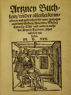
The first book focused solely on dentistry was the "ArtzneyBuchlein" in 1530 and the first dental textbook written in English was called "Operator for the Teeth" by Charles Allen in 1685.
 

Tuesday, September 3, 2013

Examination and Self Screening for Oral Cancer-Chart

Here is the diagrammatic presentation of  Examination and Self Screening for Oral Cancer

Examination and Self Screening for Oral Cancer-Chart

Wednesday, August 28, 2013

Herpes Simplex Infections


Herpes simplex virus
A range of infections, mainly viral, can produce oral blistering, but most patients present with ulceration after the blisters break. Herpesviruses are frequently responsible (Figure 9.1). Affected patients are largely children and there is often fever, malaise and cervical lymphadenopathy.
More severe manifestations and recalcitrant lesions are seen in immunocompromised people.


Herpes simplex
Definition: Herpes simplex virus (HSV) infection is common and affects mainly the mouth (HSV-1 or human herpesvirus-1; HHV-1), or genitals or anus (HSV-2; HHV-2). Initial oral infection presents as primary herpetic stomatitis (gingivostomatitis). All herpesvirus infections are characterized by latency (Figure 9.2), and can be reactivated. Recurrent disease usually presents as herpes labialis (cold sore).
Prevalence (approximate): Common. Age mainly affected: Herpetic stomatitis is typically a childhood infection seen between the ages of 2–4 years, but cases are increasingly seen in the mouth and/or pharynx in older patients.

Gender mainly affected: M : F.

Etiopathogenesis: HSV, a DNA virus, is contracted from infected skin, saliva or other body fluids. Most childhood infections are with HSV-1, but HSV-2 is often implicated more often at later ages, often transmitted sexually. UNC-93B1 gene mutations predispose to herpesvirus infection.

Diagnostic features
History: The incubation period is 4–7 days. Some 50% of HSV infections are subclinical and may be thought to be “teething” because of oral soreness.

Clinical features: Primary stomatitis presents with a single episode of multiple oral vesicles which may be widespread, and break down to form ulcers that are initially pinpoint but later fuse to produce irregular painful ulcers (Figure 9.3). Gingival edema, erythema and ulceration are prominent (Figure 9.4). The tongue is often coated and there may be oral malodor.
Herpetic stomatitis probably explains many instances of “teething”.

Extraoral features: Commonly include malaise, drooling, fever and cervical lymph node enlargement.
Complications of HSV infection occasionally include erythema multiforme or Bell palsy. HSV-1 appears to increase the risk of developing Alzheimer disease. Rare complications include meningitis, encephalitis and mononeuropathies, particularly in people with impaired immunity, such as infants whose immune responses are still developing, or immunocompromised patients.

Differential diagnosis: Other oral infections and leukemic gingival infiltrates.
Investigations: The diagnosis is largely clinical but blood tests to exclude leukemia (full blood picture and white cell count) may be indicated, and a rising titer of serum antibodies is diagnostically confirmatory but only retrospectively. Cytology, viral DNA sequentiation, culture, immunodetection or electron microscopy are used occasionally (Figures 9.5a–c).

Management
Treatment aims to limit the severity and duration of pain, shorten the duration of the episode, and reduce complications. Management includes a soft diet and adequate fluid intake. Antipyretics/analgesics such as paracetamol help relieve pain and fever. Products containing aspirin must not be given to children with any fever-causing illness suspected of being of viral origin, as this risks causing the serious and potentially fatal Reye syndrome (fatty liver plus encephalopathy).
Local antiseptics (0.2% aqueous chlorhexidine mouthwashes) may aid resolution. Aciclovir orally or parenterally is useful especially in immunocompromised patients. Valaciclovir or famciclovir may be needed for aciclovir-resistant infections.

Prognosis
Good, though HSV remains latent thereafter in the trigeminal ganglion and recurrences may occur.

Recurrent herpes labialis
Definition: Recurrent blistering of the lips caused by HSV reactivation. Prevalence (approximate): 5% of adults.
Age mainly affected: Adults.
Gender mainly affected: M = F.
Etiopathogenesis: HSV latent in the trigeminal ganglion travels to mucocutaneous junctions supplied by the trigeminal nerve, producing lesions on the upper or lower lip, occasionally the nares or the conjunctiva or, occasionally intraoral ulceration. Fever, sunlight, trauma, hormonal changes or immunosuppression can reactivate the virus which is shed into saliva, and there may be clinical recrudescence.

Diagnostic features
History: Oral premonitory symptoms may be tingling or itching sensation on the lip in the day or two days before, followed by appearance of macules, then papules, vesicles and pustules.
Clinical features: Oral lesions start at the mucocutaneous junction and heal usually without scarring in 7–10 days (Figure 9.6). Widespread recalcitrant lesions may appear in immunocompromised patients.
Extraoral: Occasionally lesions become superinfected with Staphylococcus or Streptococcus, resulting in impetigo. In immunocompromised persons, extensive and persistent lesions may involve the perioral skin. In atopic persons, the lesions of herpes labialis may spread widely to produce eczema herpeticum.

Differential diagnosis: Impetigo and other causes of blisters.
Investigations are rarely needed as the diagnosis is largely clinical.

Management
Penciclovir 1% cream, aciclovir 5% cream or silica gel applied in the prodrome may help abort or control lesions in healthy patients. Systemic aciclovir or other antivirals may be needed for immunocompromised patients.

Prognosis
Usually good but immunocompromised patients can develop recalcitrant lesions.

Recurrent intraoral herpes
Recurrent intraoral herpes in healthy patients tends to affect the hard palate or gingiva, as a small crop of ulcers usually over the greater palatine foramen, following local trauma (e.g. palatal local anesthetic injection), and heals within 1–2 weeks.
Recurrent intraoral herpes in immunocompromised patients may appear as chronic, often dendritic, ulcers frequently on the tongue ( herpetic geometric glossitis). Clinical diagnosis tends to underestimate the frequency of these lesions.
Management: The aims are to limit the severity and duration of pain, shorten the duration of the episode, and reduce complications. Symptomatic treatment with a soft diet and adequate fluid intake, antipyretics/analgesics (paracetamol), local antiseptics (0.2% aqueous chlorhexidine mouthwashes) usually suffices. Systemic aciclovir or other antivirals may be needed for immunocompromised patients.

Popular Posts

Join This site