Wednesday, January 4, 2012

Conventional Over-Denture-Introduction

A conventional over-denture rests over some healthy natural tooth roots.

An overview of the Jaw
Maintaining a denture on the jaw bone ridge (called alveolar ridge) is essential to preventing it from becoming loose during eating, speaking and other activities.
Preserving the alveolar ridge facilitates denture stability
The body tends to conserve energy and nutrients by maintaining only structures with apparent immediate value. A typical example is the bulk reduction of a broken leg held immobile by a full leg cast for a month or more.
The body "recognizes" the only one purpose for alveolar bone is to hold tooth roots.
Alveolar bone no longer supporting a tooth root is removed, or literally dissolved away by the body. This is called resorption or simply shrinkage.
Resorption progresses at varying rates in the same person at different times and at different rates between different people. Resorption progresses rapidly within the first year of loosing a tooth after which time the rate progresses at a slower pace.


Preserving the Jaw bone
IF the maximum amount of bone is to be maintained then preserving the maximum number of healthy tooth roots should achieve that end.

Preserving the sensation of having teeth
Studies demonstrate that even though only roots are preserved, and they are covered by a denture, a patient still has sensory input sensations similar to that experienced with teeth, as opposed to individuals with conventional dentures and no preserved roots. Over-denture patients also appear to have a more natural perceived directional sense in their chewing activities. In other words, many patients relate that they still feel like they have teeth - - a positive comment.

Why an over-denture?
If a patient is treatment planned to have a denture, and the roots of some remaining teeth are supported in healthy alveolar bone - - then a conventional over-denture is a viable consideration.
However, only a licensed dental professional can determine if a conventional over-denture is a suitable consideration for a certain person, after a comprehensive examination.

Some characteristics of a conventional over-denture
  • Most of a tooth crown (that part of the tooth above the gums) is removed. This often necessitates root canal therapy if not already done.
  • Remaining tooth, projecting above the gum, is rounded and usually covered with a similarly shaped artificial crown-like covering.
  • Various configurations and extensions may be built onto some retained roots. In those cases, that portion of the denture overlying these configurations is modified to contain attachments that clip onto a framework or receive the individual extensions. In addition to preserving alveolar bone and sensory input, the denture is securely held in place, but may be comfortably and easily removed for cleaning.
Advantages of a conventional over-denture
  1. Feels more like having teeth
  2. More retentive in many cases
  3. Helps reduce shrinkage of surrounding bone
  4. Reduces pressure to portions of the alveolar ridge
  5. Positive psychological advantage of still having teeth
Disadvantages of a conventional over-denture
  1. Scrupulous oral hygiene is essential in order prevent decay and gum disease.
  2. The over-denture may feel bulkier than a conventional denture.
  3. Frequent maintenance examinations are necessary.
  4. Generally this is a more expensive approach than a conventional denture.
Implant Over-Denture
An implant over-denture connects to cylinder-like configurations (called implants) that have been surgically implanted into jaw bone.
The denture appears like a traditional prosthesis. However, that part of the denture overlying implants is modified to retain various semi-rigid attachments that receive implant extensions projecting above the gum. This arrangement helps keep a denture securely in place while eating, speaking and during other oral activities, but still allows easy self-removal of the denture for cleaning purposes.
There are two phases to this process. The first is a surgical phase consisting of two stages, and the second is a prosthetic phase (making the implant denture). 

The surgical phase
Surgical insertion stage
Implants are completely inserted into precise preparations in jaw bone. While there are various implant configurations, they are essentially cylindrical in shape and made of pure titanium metal. After implants are inserted into jaw bone, gum tissue over the implant is closed with sutures in most cases.
While a minimum of two implants may be inserted for an acceptable outcome, a person may be treatment planned to receive three or more - - depending upon individual needs and anatomical limitations. More implants will give additional support and retention to the implant denture.

Healing and surgical exposure stage
During healing, an existing or temporary denture may continue to be worn after adjustments have been made to adapt to the surgerized site. If the existing denture cannot be altered sufficiently, a provisional prosthesis should be fabricated.
Implants are left undisturbed beneath gum tissue for at least several months as determined by the dental professional. During this time bone reorganizes and grows around the implant surface, anchoring it securely into the jaw (this is called osseointegration).
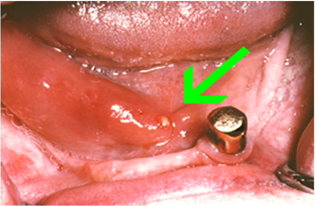
At the end of the healing stage, the top of the implant is exposed by removing gum tissue directly over it.
An extension that is then screwed into the exposed implant projects slightly above the gum tissue. After adjustments, an existing denture can be worn over an implant extension while the gum heals.
However, the denture must be reshaped to conform to surgical site contours in order to avoid unnecessary pressure areas on the newly surgerized site.

The prosthetic phase (making the implant denture)
A precision superstructure is fabricated that is screwed into the implant extensions. This superstructure may have various interface configurations ranging from interconnecting metal bars to specially shaped singular extensions.
A denture is fabricated with special provisions on the inside surface to receive various types of attachments (interlocks). Depending on the attachment, they interact in various ways with the superstructure. For example, a metal or plastic attachment may clip onto metal superstructure bars, a nylon receptacle may receive a specially configured implant extension, and so forth.
The attachment/superstructure configuration helps to securely maintain a denture while eating and speaking, and still allows a person to comfortably and easily remove the prosthesis for cleaning purposes.

Essential maintenance needs
As might be expected, exemplary oral hygiene is essential to helping prevent the development of disease around implants that could cause their failure.
Implants, superstructure, attachments and the over-denture must be checked and professionally maintained by a licensed dental professional on a regular basis. Attachments often need periodic adjustment or replacement due to wear.
While the implant over-denture approach is complex and expensive, the value received by an individual usually far exceeds monetary considerations.

How long will implant over-dentures last?
An implant may last for a lifetime (current reports show many implants lasting twenty years) or deteriorate in a few years  - many factors are involved that reduce the life expectancy of implants, such as oral hygiene, general health, habits such as smoking, grinding, and so forth. The superstructure or implant extensions may need to be replaced after five years. Depending on the implant system used, some parts may need to be replaced annually, or sooner, because of wear or deterioration. These time frames are generalities. The dental profession continues to strive for long-term durability.


Saturday, December 31, 2011

Important Post Delivary instructions following Denture Delivary

Here in this post we would like to share, Important Post delivery instructions following Denture delivery.


Congratulation! You have just received your final prosthesis. We hope you will enjoy using it. There are a few things to keep in mind:

1. You may salivate more heavily for the next several days, until your mouth is accustom to the presence of the new prosthesis.
2. You may feel awkward when talking or speaking certain words, at first. With practice, your tongue will be trained to accommodate around the prosthesis and your phonic will become normal, again. Reading out loud may help expedite the process.
3. Sore spots are normal. Please give us a call to have your prosthesis adjusted, as necessary. We want to make sure that you will be able to use your new teeth, as comfortably as possible.
4. Occasionally, due to the morphology of the underlying jaw bone, the use of adhesive cream or paste may be required to attain satisfactory retention.
5. Keep your prosthesis soak in a water bath, with denture cleansing tablet, when not in use, especially during bed time.
6. Leave your new teeth out, during bed time, allowing your gum to breath regain normal circulation.
7. Clean your denture with a toothbrush and hand/liquid soap, over a half-filled sink or bucket of water, prior to each use.
8. Avoid chewing gum or eating sticky foods.


New full or partial Denture Instructions-in Detail

Introduction
I believe that you will be very successful with your new full denture or removable partial denture. When you begin to wear your new prosthesis there is an adjustment period where your usual mouth functions may need to be relearned. These include chewing, speech, swallowing, appearance of lip posture, and ridge comfort. The following suggestions may help you in adapting to your new prosthesis and in maintaining it.

Discomfort
Avoid pain by starting with easy but nutritious food to eat. Examples of a softer diet can include fish, eggs, cottage cheese, cooked potatoes, oranges and apple sauce. If you have discomfort, remove the denture and massage the painful area with your finger. Let the gums rest and then replace the denture. Continue to use your prosthesis until your next visit. If you fail to wear the denture, no sore will be visible and precise adjustments will be very difficult.

Chewing
Try to chew with food on both sides of your mouth. If food is bilaterally placed, the denture will be less likely to tip. Try not to bite with the front teeth as this may cause the back end of the denture to move off the gums. Biting with the side teeth will give better stability. Holding the top denture up with the tongue while chewing requires talent but this habit can be very useful.

Swallowing
Pain during swallowing may simply require a minor denture base adjustment.

Saliva
With the stimulus of new dentures your mouth may have more or less saliva for a few days. Be patient and the flow will return to normal.

Speech
Speech is a very complicated and dynamic process involving all parts of the airway and mouth. Your denture has been constructed to meet the demands of stability and retention during speech. Fortunately, people are very adaptable and speech sounds very good at the time of delivery. If speech does not sound right to you, give it some time and normal body adaptation will resolve your concerns. Practice reading aloud. Do not focus undue attention on the process.

Cleaning
To remove food debris and bacterial plaque from your prosthesis, brush vigorously with a stiff denture brush. Use either soap and water, tooth paste or a commercially available denture cleaning agent. The effervescent soaking solutions are also useful. Follow the manufacturer’s instructions. Wash your denture over a basin of water or a cloth. If they are dropped on a hard surface, the acrylic portion may fracture and any metal may bend.

Sleep
In general, take the dentures out or at least remove the lower denture for the night. This will allow the gums to rest. If this causes the jaw joints to hurt replace the dentures and use your best judgment for comfortable
sleep.

Recall
Post delivery follow-up usually requires three visits. More are available as needed. After the first year, annual recall visits are useful to monitor changes in the shape of the ridges, wear of the teeth and general oral health. If there are problems with pain, chewing, or with wear or breakage of the base or teeth, please make an appointment with the office at your earliest opportunity.


Free Download Glossary of Prosthodontic Terms

Free Download Glossary of Prosthodontic
(Prosthetic Dentistry) Terms
Click Here

Thursday, December 22, 2011

Note on Necrotizing Sialometaplasia


It is spontaneous condition of an unknown cause usually of the palate in which large area of the surface epithelium underlying connective tissue and all the associated minor salivary glands become necrotic while the ducts under go squamous metaplasia. 

Clinical features:
Usually the location is at the junction of the hard and the soft palatebut it may also be present at tongue, retromolar pad and the nasal cavity.
NSM is characterized by deep seated ulceration it is punched out
With in its deep crater are the gray granular lobules which represents the necrotic minor salivary glands.
It is 2-3 cm in diameter.
It is asymptomatic but there may be numbness or burning pain.

Histopathology:
In the palatal epithelium there is no zone of ulceration which replaced by fibrin granulation tissue.

The lobules of minor salivary glands undergo coagulation necrosis.
There scattered neurophils and foamy histocytes present in zone of necrosis.

Treatment:
No treatment is required once the diagnosis is confirmed by histological examination .
The ulcer area heals by its self with in 1-3 months.
Necrotizing Sialometaplasia

Necrotizing Sialometaplasia


Wednesday, December 14, 2011

Notes on Sialolithiasis-Clinical features, Investigations, Histopathology and Treatment


Sialolithiasis
There is presence of one or more round or oval calcified structures in the duct of the major or minor salivary glands( salivary stones)

How the stone is formed:
It is assumed that mucin proteins and desquamated ductal epithelial cells form a small nidus on which the calcium salts are precipitated, this nidus then allows concentric lamellar crystallizations to occur and thus sialolith increases in size as a layer by layer gets deposited on it

Clinical features of sialolithiasis:
About 80%of sialolith affects the major salivary glands and there is more predilections for the submandibular gland.
Stones are rare in children the average age is the 4th decade with no sex preference.

They are asymptomatic discovered on dental radiographs.
If symptomatic the chief complains are pain and swelling . Swelling is results as there is ductal dilatation caused by the ductal blockade.
The pain is described as pulling drawing or stinging.


Sialolithiasis Investigations:
Panoramic radiograph.
Ultra sound imaging
orsailography

Histopathology of sialolithiasis:
Stone: On gross examination most stones are yellow or white in colour. they may be round to oval
  - some of the stones are nodular
  - after decalcification the stone shows concentric rings as of the annual rings of a tree trunk
   -The stone is acellular and amorphous in nature and may contain microbial colonies.
Ducts: the ductal lining that surrounds sialolith shows variety of reactive changes.
   - there is squamous and mucus cell
     metaplasia and changes to stratified squamous epithelium with numerous mucous goblet cells


Sialolithiasis treatment:
  • Many of the major salivary gland sialoliths can be removed by manipulation of the stone through major duct orifice
  • When manipulation fails then a surgical cut is made into the main duct
  • In triangular, or multiple stones and long standing obstructions removal of the stone and sialadenectomy is done.

Popular Posts

Join This site