Monday, March 25, 2013

RADIOLOGICAL DIFFERENTIAL DIAGNOSIS OF COMMON LESIONS IN ORO MAXILLO FACIAL REGION


1. PERIAPICAL RADIOLUCENCIES
·           Granuloma
·           Radicular cyst
·           Abscess
·           Apical scar
·           Surgical defect
·           Periodontal disease
·           Chronic suppurative osteomyelitis
·           Periapical cemental dysplasia (osteolytic stage)
·           Cementoblastoma (osteolytic stage)
·           Cementifying and ossifying fibromas (osteolytic stage)
·           Odontogenic and nonodontogenic cysts
·           Odontogenic and nonodontogenic benign tumors
·           Malignant tumors

2. PERIAPICAL RADIOPACITIES
·         Periapical cemental dysplasia (calcified stage)
·         Cementoblastoma (calcified stage)
·         Cementifying and ossifying fibromas (calcified stage)
·         Condensing osteitis
·         Osteosclerosis
·         Socket sclerosis
·         Florid osseous dysplasia (diffuse cementosis)
·         Tori
·         Hypercementosis


3. PERICORONAL RADIOLUCENCIES

Do NOT contain radiopacities:
·         Follicular space
·         Dentigerous cyst
·         Unicystic (mural) ameloblastoma
·         Odontogenic keratocyst
·         Ameloblastoma
·         Ameloblastic fibroma

MAY contain radiopacities:
·         Ameloblastic fibro-odontoma
·         Adenomatoid odontogenic tumor
·         Calcifying epithelial odontogenic tumor (Pindborg tumor)
·         Calcifying odontogenic cyst (Gorlin cyst)

4. SOLITARY CYSTLIKE RADIOLUCENCIES

Normal anatomy and Variations
·         Tooth follicle (early stage)
·         Large marrow space (wide inter-trabecular space)
·         Post extraction socket
·         Surgical defect

Odontogenic cysts:
·         Primordial
·         Lateral periodontal
·         Residual
·         Odontogenic keratocyst

Fissural cysts:
·         Incisive canal (nasopalatine)
·         Globulomaxillary
·         Median palatine
·         Median mandibular

False cysts:
·         Traumatic bone cyst
·         Stafne bone cavity

Odontogenic tumors:
·         Ameloblastoma
·         Odontogenic adenomatoid tumor
·         Calcified epithelial odontogenic tumor(Pindborg tumor)
·         Ameloblastic fibroma
·         Cementifying and ossifying fibromas (osteolytic stage)

Giant cell and nonodontogenic tumors:
·         Central giant cell granuloma
·         Giant cell lesion of hyperparathyroidism
·         Non-odontogenic benign tumors

5. SOLITARY RADIOLUCENCIES WITH DIFFUSE IRREGULAR BORDERS
·         Chronic alveolar abscess
·         Chronic osteomyelitis
·         Osteoradionecrosis
·         Squamous cell carcinoma
·         Metastatic tumors to the jaws
·         Fibrous dysplasia (early stage)
·         Histiocytosis X
·         Osteosarcoma and chondrosarcoma
·         Ewing's sarcoma
·         Burkitt's lymphoma

6. MULTILOCULAR RADIOLUCENCIES

Cysts:
·         Odontogenic keratocyst (basal cell nevus syndrome)
·         Aneurysmal bone cyst


Tumors:
·         Ameloblastoma
·         Odontogenic myxoma
·         Central hemangioma of bone
·         Metastatic tumors to the jaws

Others:
·         Cherubism
·         Central giant cell granuloma
·         Giant cell lesion of hyperparathyroidism

7. SOLITARY RADIOPACITIES
·         Exostoses and tori
·         Osteoma
·         Retained root
·         Osteosclerosis
·         Socket sclerosis
·         Mature cementoma
·         Complex composite odontoma
·         Compound composite odontoma
·         Calcifications in soft tissue
·         Foreign objects in the jaws
·         Fibrous dysplasia
·         Garré's osteomyelitis

8. MIXED RADIOLUCENT - RADIOPAQUE LESIONS

Inflammatory:
·         Condensing osteitis

Osteomyelitis:
·         Chronic suppurative osteomyelitis
·         Tuberculous osteomyelitis
·         Syphilitic osteomyelitis
·         Actinomycotic osteomyelitis
·         Osteoradionecrosis

Cementomas (intermediate and calcified stages):
·         Periapical cemental dysplasia
·         Cementifying and ossifying fibroma
·         Cementoblastoma
·         Florid osseous dysplasia (diffuse cementosis)

Others:
·         Fibrous dysplasia
·         Paget's disease
·         Osteosarcoma
·         Chondrosarcoma
·         Osteoblastic metastatic carcinoma

Pericoronal mixed lesions:
·         Ameloblastic fibro-odontoma
·         Odontogenic adenomatoid tumor
·         Calcifying epithelial odontogenic tumor (Pindborg tumor)
·         Calcifying odontogenic cyst (Gorlin cyst)

9. GENERALIZED RADIOPACITIES
·         Florid osseous dysplasia (diffuse cementosis, sclerotic cemental masses)
·         Paget's disease
·         Osteopetrosis
·         Gardner's syndrome
·         Infantile cortical hyperostosis
·         Albright’s syndrome

Thursday, March 21, 2013

SOFT TISSUE CALCIFICATIONS IN ORO-MAXILLO FACIAL REGION-with Radiographs


Pathologic calcification of soft tissues occurs when calcium and other mineral salts are deposited in a tissue or in a passage. There are three types of pathologic calcifications: 1) Dystrophic calcification is that which occurs in degenerating and dead tissues. Calcificationof the larval stage of tapeworm (cysticercus) is an example of dystrophic calcification. 2)Metastatic calcification is that in which calcium (and other) salts are deposited in previouslyundamaged tissue as a result of an excess of salts in the circulating blood.Hyperparathyroidism is an example of metastatic calcification which occurs in kidneys andblood vessels. 3) Calcinosis is calcification that occurs in or under the skin. Scleroderma,myositis ossificans, and multiple miliary osteomas are examples of calcinosis.
SIALOLITH
A sialolith is a stone (salivary calculus) within a salivary gland or duct. The formation of a sialolith is called sialolithiasis and occurs as a result of precipitation of calcium andphosphate salts around a nidus of mucous or bacterial debris. Sialoliths occur as single or multiple stones and can cause swelling and pain. The pain is experienced during salivary stimulation and is intensified at mealtimes. The accumulation of saliva in the gland produces swelling and the gland becomes enlarged and firm. The pain is produced as a result of the buildup of pressure due to the accumulation of saliva behind the stone. The pain gradually subsides and the swelling diminishes because the stone usually does not completely block the flow of saliva. If a sialolith is small or does not obstruct the flow of saliva, there may be an absence of pain and swelling. Most stones are found in the submandibular duct (Wharton's) and gland than in the parotid duct (Stensen's) and gland because of the viscous consistency and mineral content of the saliva from the submandibular gland and the long, irregular length of the Wharton's duct.
The best radiographic projection for visualizing sialoliths in the submandibular duct and gland is the standard mandibular occlusal view. Occasionally, sialoliths are seen incidentally on periapical radiographs, in which case they may be misdiagnosed as osteosclerosis. To differentiate a sialolith from an osteosclerosis, use the Clark's rule of tube-shift technique to localize objects, that is, to find the bucco-lingual relationship. Stones in the parotid duct and gland are best demonstrated by placing a periapical film in the buccal vestibule and x-radiating them with a reduced exposure time. Approximately 20% to 40% of all sialoliths are radiolucent. When this is suspected, sialography (injection of a radiopaque dye into the ductal opening and then x-radiating them) must be undertaken to visualize the stones. The duct or gland injected with the radiopaque dye shows the radiolucent sialolith as a non-filling defect. A sialolith must be differentiated from other soft tissue calcifications, especially from a calcified lymph node. The latter is usually asymptomatic and sialography may be required to distinguish the two lesions.
Fig 11-1 Mandibular occlusal projection shows a sialolith (salivary calculus) in the duct of the submandibular gland (Wharton's duct). The patient has a history of pain and swelling in the salivary gland which is intensified at mealtime when saliva flow is stimulated. The pain gradually subsides and swelling diminishes because the stone usually does not completely block the flow of saliva.
Fig. 11-2 On periapical radiographs, the radiopacity may be misdiagnosed as osteosclerosis. To differentiate an osteosclerosis from a sialolith, take two radiographs using different vertical (or horizontal) angulations of the x-ray beam. If the radiopacity changes its position in relation to the adjoining teeth, as shown here, the radiopacity is a sialolith in the floor of the oral cavity (Clark's rule: same lingual, opposite buccal). Another method to identify a submandibular sialolith is to take an occlusal projection.
Fig. 11-3 A sialolith on a panoramic radiograph may be misdiagnosed as a calcified lymph node. In the absence of clinical signs and symptoms it is difficult to differentiate the two types of calcifications unless a sialogram is made.
Fig. 11-4 Sialogram showing an obstruction in the Wharton's duct preventing the flow of the radiopaque dye into the submandibular salivary gland. The stone (arrow) is blended with the radiopaque dye.

CALCIFIED LYMPH NODE
A calcified lymph node is indicative of a prior chronic infection involving the node. A history of successfully treated tuberculosis is often associated with this calcification. The condition is asymptomatic. It may involve a single node or a chain of submandibular or cervical nodes. The calcified superficial lymph nodes are palpable as bony, hard, round or linear masses with variable mobility. They are often observed on a panoramic radiograph, where they may appear below the inferior border of the mandible and near the angle of the mandible. Calcified lymph nodes are often found incidentally on radiographic examinationsSome may be radiographically projected over the mandibular bone and may be misdiagnosed as osseous lesions.
A calcified submandibular lymph node may be difficult to distinguish from a sialolith. The former is invariably asymptomatic whereas the latter is frequently accompanied by pain and swelling at mealtimes. Sialography may be required to distinguish the two lesions.
Fig. 11-5 Calcified lymph nodes located inferior to the angle of the mandible. Priorchronic infection of the lymph nodes may result in calcification of the nodes. A history of successfully treated tuberculosis is often associated with this calcification. This asymptomatic condition may involve a single node or a chain of nodes.
Fig. 11-6 A lateral cervical radiograph shows a chain of calcified lymph nodes.
PHLEBOLITH
Phleboliths are calcified thrombi that occur in veins or sinusoidal vessels of hemangiomas involving the soft tissues adjacent to the jaws. On a radiograph they appear as round or oval bodies which may exhibit concentric calcific rings similar to the cross section of an onion. Phleboliths may occur singly or as multiple calcifications. On periapical radiographs, calcifications may be superimposed on the mandible and thus misdiagnosed as osseous lesions within the jaw or as sialoliths.
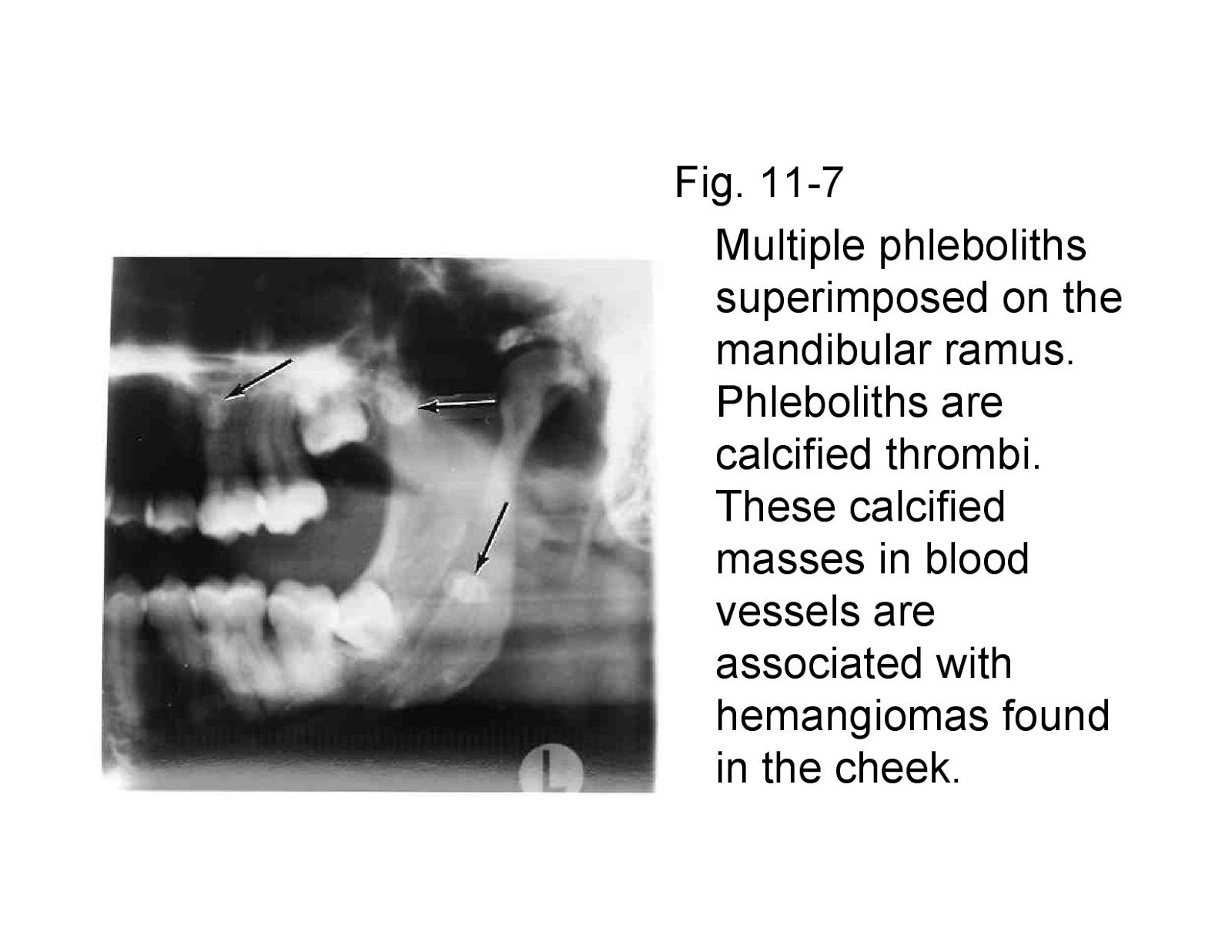
Fig. 11-7 Multiple phleboliths superimposed on the mandibular ramus. Phleboliths are calcified thrombi. These calcified masses in blood vessels are associated with hemangiomas found in the cheek.
Fig. 11-8 Multiple phleboliths of various sizes in cavernous hemangioma of the face. The radiograph is of the patient's cheek.

CALCIFICATION OF ARTERIES
Calcification of the walls of arteries occurs in arteriosclerosis and in secondary inflammatory conditions involving arteries. The calcium salts are deposited within the medial coat of the vessels. Calcification can occur in a number of arteries of the body (iliac, femoral, abdominal aorta, etc.), however, in the facial region the facial artery is the one that is often involved. Calcified arteries of the cheek and oral cavity may appear as faint images on periapical radiographs. In the Sturge-Weber syndrome (capillary hemangiomas of the face, oral mucosa, and cranium), the cranial hemangiomas often show marked calcification of the blood vessels.
Fig. 11-9 Calcification of the facial artery. It may occur in arteriosclerosis and represents an inflammatory process.
Fig. 11-10 Calcification of the facial artery. The radiopacity of the artery is the result of deposition of calcium salts within the medial coat of the vessel.

ANTROLITH
A calcified mass in the maxillary sinus is called an antrolith. It is produced by calcification of a nidus which may be a bone chip, root fragment, foreign object, or stagnant mucus in sites of previous inflammation. Most of the antroliths are asymptomatic and are detected incidentally on radiographic examinations. However, on rare occasions when an antrolith continues to grow and become very large, it may be associated with sinusitis. Antroliths must be differentiated from root fragments in the maxillary sinus. A root fragment will show the root anatomy such as the presence of a pulp canal in a cone-shaped (root-shaped) radiopacity. When calcification comparable to an antrolith occurs in the nasal fossa, it is called a rhinolith.
Fig. 11-11 Calcified mass in the maxillary sinus is called an antrolith. A foreign object, bone chip, root fragment or stagnant mucus acts as a nidus for calcific deposits. It is usually asymptomatic.
Fig. 11-12 Antrolith (stone in maxillary sinus) on the floor of the sinus. It is asymptomatic.

MULTIPLE MILLIARY OSTEOMAS OF SKIN
(Osteoma cutis, calcinosis cutis)
Multiple milliary osteomas of skin, also known as calcinosis cutis, are situated in the cutis and subcutis. Some of these calcifications are associated with acne or some other form of dermatosis. They are found incidentally on radiographic examinations. They appear as doughnut-shaped radiopacities with radiolucent centers which represent the central marrow cavities. Multiple miliary osteomas are imaged better by placing a dental film in the vestibules and against the inside surface of the cheek and using a reduced exposure time.
Fig. 11-13 Multiple miliary osteomas of skin are soft tissue calcifications of skin. Some are reported to be associated with acne or some other form of dermatosis.
Fig. 11-14 Calcinosis cutis showing doughnut-shaped radiopacities.

CALCIFIED STYLOHYOID LIGAMENT AND EAGLE'S SYNDROME
Calcification of the stylohyoid ligament may sometimes be found incidentally on a panoramic radiograph and located posterior to the ramus of the mandible. It may occur unilaterally or bilaterally. In about 50% of the cases, the individuals are asymptomatic. In those cases associated with pain and discomfort, the entity is called "Eagle's syndrome".
The syndrome includes vague pain on mandibular movements such as swallowing (dysphagia), turning the head or opening the mouth, sensation of foreign body in throat, and constant dull ache in the throat. Other symptoms include headache, earache (otalgia), dizziness, pain in temporomandibular joint area and also in the base of the tongue or transient syncope. The symptoms are probably caused by the elongated styloid process impinging on the glossopharyngeal nerve. When the jaws are closed, the pain subsides in some of the cases. It is important for the dentist to be aware that pain associated with calcified stylohyoid ligament may simulate pain associated with that of the temporomandibular joint.
On a radiograph, the calcified stylohyoid ligament appears as a thin, long, tapering radiopaque process extending downwards from the styloid process. Sometimes it may extend up to the lesser horn of the hyoid bone. The farther the mineralized ligament extends towards the hyoid bone, the more likely it is that it will be interrupted by radiolucent joint like junctions. Surgical resection is required in patients exhibiting symptoms.
Fig. 11-15A Patient with Eagle’s syndrome. The stylohyoid ligaments are bilaterally calcified. Patient complained of constant dull ache in the throat, pain on turning the head, and pain in the vicinity of the temporomandibular joints.
Fig. 11-15B Calcified stylohyoid ligament. Sometimes, this calcification may be associated with Eagle's syndrome. The syndrome produces cervical pain on turning the head, upon swallowing and on opening the mouth. The patient may have headaches, and dizziness.
Fig. 11-16 Calcified stylohyoid ligament in an asymptomatic patient.
Fig. 11-17 Bilateral calcified stylohyoid ligament in a case associated with Eagle's syndrome.

CALCIFIED THYROID CARTILAGE
Calcification of the thyroid cartilage is normal and increases with age. The thyroid and cricoid cartilages have been found to undergo a greater frequency of calcification in the female population, but a higher degree of ossification has been noted in male subjects. (In the hyaline cartilages of the larynx, calcification does not always precede ossification and there is little correlation between the two).
Fig. 11-18 Calcification of the thyroid cartilage. It is asymptomatic.

MYOSITIS OSSIFICANS
In myositis ossificans, bony structures such as lamellae, lacunae and marrow are deposited in soft tissue. The cause of this ossification in muscles, ligaments, tendons and fascia is unknown. It may be caused by trauma or heavy muscular strain that occurs in certain
occupations and sports. During the healing process, the blood in the traumatized region gets organized and later calcified. The calcification takes place in the connective tissues around the muscles. The digastric, masseter, temporal and sternomastoid are usually involved. The patient finds it difficult to open the jaws when the muscles of mastication are involved. The characteristic radiographic appearance is that of strand-like calcifications along the long axis of the muscle fibers.

CYSTICERCOSIS
Cysticercosis is a helminthic (parasitic worms) disease which completes the larval phase of its life cycle in the pig. When an individual ingests eggs of the (pork) tapeworms from contaminated water or food, the larval form of the tapeworms are hatched in the gastrointestinal tract, and enter the vascular and lymphatic systems. They are then deposited in various tissues and organs of the body. At this stage, there is no radiographic evidence of their presence. After their death, the larval spaces are filled with fibrous tissue which later becomes calcified. These calcifications in muscle and subcutaneous tissue are visible on a radiograph as multiple radiopaque ovoid or elliptical objects.
Fig. 11-19 Radiograph of patient with cysticercosis. The calcified encysted larvae are seen in the soft tissues at the back of the neck and a single one is seen in the area of Wharton’s duct.

Osteomyelitis-with Radiological features

OSTEOMYELITIS

  • Suppurative osteomyelitis
  • Garrés osteomyelitis (periostitis ossificans, proliferative osteomyelitis)
  • Tuberculous osteomyelitis
  • Syphilitic osteomyelitis
  • Actinomycotic osteomyelitis
  • Osteoradionecrosis
SUPPURATIVE OSTEOMYELITIS
Osteomyelitis is an inflammatory reaction of bone to infection which originates from either a tooth, fracture site, soft tissue wound or surgery site. The dental infection may be from a root canal, a periodontal ligament or an extraction site. Suppurative osteomyelitis can involve all three components of bone: periosteum, cortex, and marrow. Usually there is an underlying predisposing factor like malnutrition, alcoholism, diabetes, leukemia or anemia.
Other predisposing factors are those that are characterized by the formation of avascular bone for example, therapeutically irradiated bone, osteopetrosis, Paget's disease, and florid osseous dysplasia. Osteomyelitis is more commonly observed in the mandible because of its poor blood supply as compared to the maxilla, and also because the dense mandibular cortical bone is more prone to damage and, therefore, to infection at the time of tooth extraction.
Acute osteomyelitis is similar to an acute primary abscess in that the onset and course may be so rapid that bone resorption does not occur and, thus, a radiolucency may not be present on a radiograph. Clinical features include pain, pyrexia, painful lymphadenopathy, leukocytosis, and other signs and symptoms of acute infection. Later, after approximately two weeks, as the lesion progresses into the chronic stage, enough bone resorption takes place to show radiographic mottling and blurring of bone. A sclerosed border called an involucrum forms around the affected area. The involucrum prevents blood supply from reaching the affected part. This results in the formation of pieces of sequestra or necrotic bone surrounded by pus. A fistulous tract may develop by the suppuration perforating the cortical bone and periosteum. The fistulous tract discharges pus onto the overlying skin or mucosa.
The radiopacity of the sequestra and the radiolucency of the pus give rise to the characteristic "worm-eaten" radiographic appearance. Radiographs also aid in locating the original site of infection such as an infected tooth, a fracture, or infected sinus.

Fig. 13-1 Chronic suppurative osteomyelitis of dental origin. The lesion discharged pus into the oral cavity. Note the radiopaque sequestra (arrow) surrounded by the radiolucent suppuration.

Fig. 13-2 Chronic suppurative osteomyelitis demonstrating a worm-eaten appearance of the body of the mandible. Note the radiopaque sequestra surrounded by the radiolucent suppuration and a radiopaque involucrum. The patient had fetid breath.
Fig. 13-3 Chronic suppurative osteomyelitis of dental origin. The radiopaque sequestrum (arrow) is surrounded by the radiolucent suppuration.
Fig. 13-4 Sequestrum that has floated into the soft tissues. Patient gave a history of a problematic tooth extraction several years ago which resulted in clinical complications.

GARRÉS OSTEOMYELITIS
(Periostitis ossificans, Osteomyelitis with proliferative periostitis) Garrés osteomyelitis or proliferative periostitis is a type of chronic osteomyelitis which is nonsuppurative. It occurs almost exclusively in children and young adults who present symptoms related to a carious tooth. The process arises secondary to a low-grade chronic infection, usually from the apex of a carious mandibular first molar. The infection spreads towards the surface of the bone, resulting in inflammation of the periosteum and deposition of new bone underneath the periosteum. This peripheral formation of reactive bone results in localized periosteal thickening. The inferior border of the mandible below the carious first molar is the most frequent site for the hard nontender expansion of cortical bone. On an occlusal view radiograph, the deposition of new bone produces an "onion-skin" appearance.
Fig. 13-5 Garrés osteomyelitis (proliferative periostitis) demonstrating an expansion of the inferior border of the mandible (onion-skin appearance) caused by the periapical infection of the mandibular first molar.
Fig. 13-6 An occlusal radiograph of Garrés osteomyelitis showing the buccal expansion of the mandible caused by infection around the root tip of the extracted first molar.
Fig. 13-7 Garrés osteomyelitis (periostitis ossificans) exhibiting localized periosteal thickening. The source of infection is not known; it could have been from an exfoliated deciduous molar tooth.

TUBERCULOSIS OSTEOMYELITIS
Tuberculosis is a chronic granulomatous disease which may affect any organ, although in man the lung is the major seat of the disease and is the usual portal through which infection reaches other organs. The microorganisms may spread by either the bloodstream or the lymphatics. Oral manifestations of tuberculosis are extremely rare and are usually secondary to primary lesions in other parts of the body. Infection of the socket after tooth extraction can also be the mode of entry into the bone by Mycobacterium tuberculosis.
Mandible and maxilla are less commonly affected than long bones and vertebrae. On a radiograph, the appearance of bony lesions is similar to that of chronic suppurative osteomyelitis ("worm- eaten" appearance) with fistulae formation through which small sequestra are exuded. Periostitis ossificans (proliferative periostitis) can also occur and change the contour of bone. Calcification of lymph nodes is a characteristic sign of tuberculosis.
Fig. 13-8 Tuberculous osteomyelitis showing the "worm-eaten" appearance similar to that of a chronic suppurative osteomyelitis
Fig. 13-9 Calcified tuberculous lymph nodes

SYPHILITIC OSTEOMYELITIS
Syphilis is a chronic granulomatous disease which is caused by the spirochete Treponema pallidum. It is a contagious venereal disease which leads to many structural and cutaneous lesions. Acquired syphilis is transmitted by direct contact whereas congenital syphilis is transmitted in utero. In congenital syphilis, the teeth are hypoplastic, that is, the maxillary incisors have screwdriver-shaped crowns with notched incisal edges (Hutchinson's teeth) and the molars have irregular mass of globules instead of well-formed cusps ("mulberry molars"). Also, a depressed nasal bridge or saddleback nose occurs because of gummatous destruction of the nasal bones.
Acquired syphilis, if untreated, has three distinct stages. The primary stage develops after a couple of weeks of exposure and consists of chancres on the lips, tongue, palate, oral mucosa, penis, vagina, cervix or anus. These chancres are contagious on direct contact with them. The secondary stage begins 5 to 10 weeks after the occurrence of chancres and consists of diffuse eruptions on skin and mucous membrane. This rash may be accompanied by swollen lymph nodes throughout the body, a sore throat, weight loss, malaise, headache and loss of hair. The secondary stage can also damage the eyes, liver, kidneys and other organs. The tertiary-stage lesions may not appear for several years to decades after the onset of the disease. In this stage of osteomyelitis, the bone, skin, mucous membrane, and liver show gummatous destruction which is a soft, gummy tumor that resembles granulation tissue. Paralysis and dementia can also occur. In the oral cavity, the hard palate is frequently involved resulting in its perforation. The gummatous destruction is painless. Syphilitic osteomyelitis of the jaws is difficult to distinguish from chronic suppurative osteomyelitis since their radiographic appearances are similar.
Fig.13-10 Syphilitic osteomyelitis of the palate. The gummatous destruction has produced a palatal perforation.
Fig.13-11 Radiograph of syphilitic osteomyelitis of the palate. The perforation which is the site of gumma of the hard palate produces a radiolucency which may be mistaken for a median palatine cyst.

ACTINOMYCOTIC OSTEOMYELITIS
Like tuberculosis and syphilis, actinomycosis is a chronic granulomatous disease. It can occur anywhere in the body, but two-thirds of all cases occur in the cervicofacial region. The disease is caused by bacteria-like fungus called Actinomyces israeli. These microorganisms occur as normal flora of the oral cavity, and appear to become pathogenic only after entrance through previously seated defects. The portal of entry for the microorganisms is either through the socket of an extracted tooth, a traumatized mucous membrane, a periodontal pocket, the pulp of a carious tooth or a fracture. In cervicofacial actinomycosis, the patient exhibits swelling, pain, fever and trismus. The lesion may remain localized in the soft tissues or invade the jaw bones. If the lesion progresses slowly, little suppuration takes place; however, if it breaks down, abscesses are formed that discharge pus containing yellow granules ( (nicknamed sulfur granules) through multiple sinuses.
There is no characteristic radiographic appearance. In some cases the lesion resembles a periapical radiolucent lesion. The more aggressive lesion resembles chronic suppurative osteomyelitis. In chronic suppurative osteomyelitis there is usually a single sinus through which pus exudes; however, in actinomycotic osteomyelitis there are many sinuses through which pus and "sulfur granules" exude.
Fig.13-12 Actinomycotic lesion similar to radicular cyst. This is not a typical appearance.

OSTEORADIONECROSIS (and effects of irradiation on developing teeth)
In therapeutic radiation for carcinomas of the head and neck, the jaws are subjected to high exposure doses of ionizing radiation (average of 5000 R). This results in decreased vascularity of bone and makes them susceptible to infection and traumatic injury. Infection may occur in irradiated bone from poor oral hygiene, extraction wound, periodontitis, denture sores, pulpal infection or dental treatment. It is therefore advisable that a patient scheduled to undergo therapeutic radiation be given dental treatment prior to radiation therapy and that after radiation therapy the patient be taught to maintain good oral hygiene.
When infection occurs in irradiated bone, it results in a condition called osteoradionecrosis which is similar to chronic suppurative osteomyelitis. The mandible is affected more commonly than the more vascular maxilla. Therapeutic radiation may affect the salivary glands, producing decreased salivation. The resulting temporary or permanent xerostomia is responsible for radiation caries of teeth and erythema of the mucosa.
A radiograph of osteoradionecrosis, shows radiopaque sequestra and surrounding radiolucent purulency similar to that of chronic suppurative osteomyelitis. The two cannot be differentiated radiographically except by the history of therapeutic radiation. Effects of irradiation on developing teeth depends on the stage of development when irradiation occurs and on the dosage administered. The injured tooth germs may either fail to form teeth (anodontia), exhibit dwarf-teeth, produce agenesis of roots, shortening and tapering of roots, or develop into hypoplastic teeth. The eruption of teeth may be retarded and their sequencing may be disturbed. Other radiation induced effect may include maxillary and/or mandibular hypoplasia.
Fig.13-13 Occlusal projection of anterior region of mandible showing osteoradionecrosis. Notice the destruction of the trabecular pattern of bone.
Fig.13-14 Osteoradionecrosis of left mandible showing the radiopaque sequestra.
Fig.13-15 Osteoradionecrosis of left mandible has resulted in a pathologic fracture.
Fig.13-16 Dwarfing of teeth as a consequence of radiation therapy

Popular Posts

Join This site